🧬 Biologie
Cellule, biologie moléculaire, génétique, systèmes physiologiques et évolution
  <div class="prog-meta">
    <span>📄 18 chapitres</span>
    <span>🖼️ 81 illustrations</span>
    <span>📖 Adapté de <a href="https://mcat-review.org/biology.php" target="_blank" rel="noopener">mcat-review.org</a></span>
  </div>

  <div class="toc">
    <h2>Sommaire</h2>
    <ol class="toc-list">
<li><span class="toc-num">01</span><a href="#ch-1">Systèmes circulatoire, lymphatique et immunitaire</a></li> <div class="chapter" id="ch-1">
<div class="chapter-header" onclick="toggleChapter(this)">
<div>
<div class="chapter-num">Chapitre 01</div>
<div class="chapter-title">Systèmes circulatoire, lymphatique et immunitaire</div>
</div>
<div class="chapter-toggle open">▼</div>
</div>
<div class="chapter-body open">
<div class="fiche-section">
<div class="fiche-section-title">Système circulatoire</div>
<ul class="fiche-list">- Oxygen delivery to tissus
- diffuses into the blood in alvéolaire (poumon) capillaires
- binds to hemoglobin in globules rouges
- gets transported to tissus
- used in respiration cellulaire
- Carbon dioxide delivered out
- respiration cellulaire makes CO2: carbonic anhydrase converts it to bicarbonate.
- CO2 gets transported by blood: dissolved CO2, dissolved bicarbonate ion (major), bound to hemoglobin and protéines plasmatiques
- diffuses out of the alvéolaire capillaires
- exhaled out
- Nutriments
- nutriments absorbed (either by diffusion or transport actif) into flux sanguin in the intestin grêle.
- nutriments can also be released into the flux sanguin by cellules. For example, glucagon causes glucose to be released into the flux sanguin.
- nutriments can be taken up by cellules. For example, insulin causes cellules to take in glucose from blood.
- Hormones released by glandes endocrines, circulate the blood in order to reach their cellules cibles.
- Fluids and ions circulate the blood and are regulated by how much reabsorption of water and salt occurs in the rein.
- Urea = déchet métabolique, travels in the blood to the reins, where it is filtered out and passed in urine.
- Role in thermorégulation
- Vasoconstriction conserves heat. When it's cold, vasoconstriction occurs in the artérioles that feed the skin. Less flux sanguins near the surface of the skin, less heat lost.
- Vasodilatation cools you down. When it's hot, vasodilatation occurs in the artérioles that feed the skin. More skin flux sanguin, more heat lost to the surroundings.
- Four-chambered heart (structure, function)
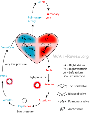
- Deoxygenated blood returns to the heart: superior/inferior vena cava → right atrium
- Deoxygenated blood gets pumped to the poumons: right atrium → right ventricle → pulmonary artère → poumons
- Blood arrives at the poumons and gets oxygenated.
- Oxygenated blood returns to the heart: poumons → pulmonary veine → left atrium
- Oxygenated blood gets pumped to the body: left atrium → left ventricle → aorte
- Blood going through the heart including the valves
- Vena cava
- Right atrium
- Tricuspid valve
- Right ventricle
- Pulmonary valve
- Pulmonary artère
- Poumon
- Pulmonary veine
- Left atrium
- Bicuspid (Mitral) valve
- Left ventricle
- Aortic valve
- Aorte
- Systolic and diastolic pressure
- pression artérielle = pressure blood exert on the walls of the vaisseau sanguin.
- systolic pressure = pression artérielle when blood is being pumped (the ventricles are contracting).
- diastolic pressure = pression artérielle when blood is not being pumped (the ventricles are relaxing).
- Pulmonary and systemic circulation
- Pulmonary circulation = heart → poumons → back to heart = oxygenates blood
- Systemic circulation = heart → body → back to heart = delivers oxygenated blood to body
- Pulmonary circulation = shorter than systemic circulation = less resistance = less pression artérielle.
- Systemic circulation: vasodilatation when niveaux d'oxygène are low → more flux sanguin to oxygen-starved tissu.
- Pulmonary circulation: vasoconstriction when niveaux d'oxygène are low → less flux sanguin to low oxygen/blocked alvéoles → more flux sanguin to good alvéoles where échange gazeux can occur.
- Arterial and venous systems (artères, artérioles, veinules, veines)
- structural and functional differences
- Blood flows from artère → artériole → capillaire → veinule → veine.
- Artère
- Elastic artère
- Aorte and its major branches.
- Major function = provide elastic pipe for blood straight out of the heart.
- Lots of elastic tissu.
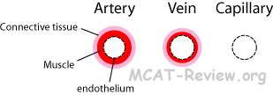
- Layers: endothelium, muscle lisse, tissu conjonctif.
- Not active in vasoconstriction.
- Muscular (distributing) artères
- Major function = distribute blood to specific organes.
- Lots of muscle.
- Layers: endothelium, lots of muscle lisse, tissu conjonctif.
- Some activity in vasoconstriction.
- Artériole
- Ranges from being like a smaller version of the artère, to being a larger version of the capillaire with muscles lisses spiralling around it.
- Major function = controls flux sanguin to the capillaires.
- Active in vasoconstriction. The artérioles allow the body to control which tissus gets more blood.
- The artériole is the most important site for vasoconstriction. Although other vessels are capable of vasoconstriction, you should always think of the artériole when you see vasoconstriction.
- Capillaire
- Layer: single cellule thick endothelium.
- Major function: blood-tissu solute exchange.
- Not active in vasoconstriction.
- Veinule
- Ranges from being like a large capillaire to being like a small veine.
- Major function: merge of capillaires to be conducted to veines.
- No vasoconstriction.
- Veine
- Layers: endothelium, muscle lisse, tissu conjonctif.
- Major function: returns blood back to the heart.
- Has valves to prevent the back flow of blood.
- Breathing, muscles squelettiques, and muscle lisse adaptations help flux sanguin through the veine at low pressure.
- Vasoconstriction can occur in the veine.
- You can argue that the aorte has a single aortic valve right where it connects to the heart. But for the purposes of the MCAT, artères don't have valves, veines do.
- Thickness: artère > veine > artériole > veinule > capillaire
- Differences between artères and veines
- artères are thicker, more muscular than veines.
- veines have valves, artères don't.
- artères carry blood away from the heart (oxygenated except for pulmonary artère). Veines carry blood back into the heart (deoxygenated except for pulmonary veine).
- Differences between artère and artériole
- artérioles are smaller.
- vasoconstriction occurs predominantly at the artérioles.
- pressure and flow characteristics
- Blood pressure of artères > artérioles > capillaires > veinules > veines
- Blood pressure is highest in the artères (specifically the aorte) because the heart pumps directly into the aorte.
- Blood pressure is lowest in the veines (specifically the vena cava) because flow resistance brings the pressure down.
- Blood pressure is also lower when you elevate a vaisseau sanguin (think physics, P = ρgh, where h is the depth - raising your arm like taking it to shallower water)
- Blood pressure can be regulated by vasoconstriction (increase bp), vasodilatation (decrease bp), and hormones (ADH, aldosterone, renin, adrenaline all increases bp).
- Blood flows from artère → artériole → capillaire → veinule → veine.
- Blood squirts from artères, flows from veines, and oozes from capillaires.
- The élasticité of artères causes blood to flow even when the heart is resting between pumps (this is why your diastolic pression artérielle is not zero)
- Adaptations that help flux sanguin through the veine at low pressure:
- Respiratory pump: when you inhale, your estomac squeezes on the veines, and your chest sucks on it.
- Muscular pump: muscle squelettique squeezes on the veines when you exercise.
- When you're scared, muscles lisses around veines constrict and squeezes blood.
- Capillaire beds
- mechanisms of gas and solute exchange
- Diffusion is the major mechanism of gas and solute exchange, whether it is diffusion as a free molecule, or bound to carrier protéines.
- Continuous capillaire
- No pores on endothelial cellules. May have clefts at cellule boundaries.
- Exchange may occur through the clefts, or by vesicle trafficking through endothelial cellules.
- Found in skin and muscles.
- Blood-cerveau barrier = sealing of clefts by jonctions serrées.
- Fenestrated capillaire
- Small pores, large enough for molecules, but not cellules sanguines to leak through.
- Found in intestin grêle to facilitate nutrient absorption.
- Found in organes endocriniens to allow passage of hormones.
- Found in reins to allow blood filtration.
- Sinusoidal capillaire
- Large pores, large enough for cellules sanguines to leak through.
- Found in tissus lymphoïdes, foie, rate, moelle osseuse.
- Large pores facilitate lymphocyte travel to tissus.
- Large pores also facilitate cellule sanguine modifications.
- mechanism of heat exchange
- rayonnement - your body gives off IR signal.
- conduction - you touch something cold, or take a hot bath.
- evaporative cooling - you sweat, and it cools you as it evaporates.
- source of peripheral resistance (no longer tested)
- Blood viscosité: cellules sanguines and protéines plasmatiques give blood a higher resistance to flow compared to water. Diseases that increase the amount of cellules sanguines increase resistance.
- Total vaisseau sanguin length: more vaisseaux sanguins you have, the more resistance to flow. Overweight = more vaisseaux sanguins to service the fat cellules = more resistance.
- Blood vessel diameter: vasoconstriction increases resistance, vasodilatation decreases it. Obstruction from plaques inside vaisseaux sanguins also increases resistance.
- Composition of blood
- plasma, chemicals, cellules sanguines
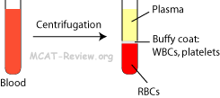
- plasma = water and chemicals = mostly water, protéines plasmatiques, electrolytes, gases, nutriments, wastes, hormones.
- cellules sanguines
- globules rouges (RBCs or erythrocytes)
- contain hemoglobin, transports O2 and CO2
- no noyau, which gives it a biconcave disk shape
- most abundant cellule in blood.
- globules blancs (WBCs or leukocytes)
- larger than RBCs
- lobed or irregular shaped nuclei
- fights off agents pathogènes
- platelets
- technically not cellules, but cellule fragments
- responsible for clotting blood
- erythrocyte production and destruction (rate, moelle osseuse)
- Bone marrow = makes RBCs from cellules souches.
- Rate = destroys aged and damaged RBCs.
- Other sites for RBC destruction include the foie and moelle osseuse.
- Components of hemoglobin from destroyed RBC gets recycled
- iron = recycled
- heme → bilirubin → bile → excreted in feces
- protéine (globin) = broken down to acides aminés
- régulation of plasma volume
- Blood osmolarity
- Higher osmolarité sanguine → water goes into blood → higher volume sanguin
- Lower osmolarité sanguine → water goes into tissus → lower volume sanguin
- ADH (vasopressin): ↑ réabsorption d'eau in rein.
- Aldosterone: ↑ réabsorption de sel, leads to ↑ réabsorption d'eau in rein.
- coagulation, mécanisme de coagulations, role of foie in production of facteurs de coagulation
- Platelets contain enzymes and chemicals needed involved in the processus de coagulation.
- Foie produces facteurs de coagulation (eg. fibrinogen), which circulates in plasma sanguin.
- Coagulation = liquid blood → gel
- Clotting mechanism:
- Platelet plug formation: wound + platelets → platelets clump at wound, release chemicals, activates facteurs de coagulation.
- Coagulation: series of facteur de coagulation/enzyme activation that ends in fibrinogen → fibrin. Fibrin being the fiber mesh that seals the clot.
- Retraction and repair: clot contracts, gets compact, but after the wounded vaisseau sanguin repairs itself, the clot dissolves.
- Oxygen and dioxyde de carbone transport by blood
- hemoglobin, hematocrit
- hemoglobin = (heme + globin) x 4
- heme = chemical ligand binding iron
- globin = protéine that surrounds heme
- 4 subunits of the heme-globin complex form a tetramer called hemoglobin.
- hemoglobin can bind oxygen and dioxyde de carbone
- hematocrit = % volume of blood that is globules rouges, usually ~ 45%
- oxygen content
- each iron atom in hemoglobin can bind one oxygen.
- hemoglobin has 4 subunits containing 4 iron atoms.
- each RBC has hundreds of millions of hemoglobin molecules.
- affinité pour l'oxygène
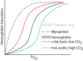
- hemoglobin has a sigmoidal liaison à l'oxygène curve. This is because liaison à l'oxygène to one subunit "relaxes" the conformation of the other subunits, and makes it easier for additional oxygen to bind.
- monoxyde de carbone binds hemoglobin tighter than oxygen.
- fetal hemoglobin binds oxygen tighter than adult hemoglobin.
- myoglobin binds oxygen tighter than hemoglobin.
- Details of oxygen transport: biochemical characteristics of hemoglobin
- modification of affinité pour l'oxygène
- Higher levels of dioxyde de carbone → lower affinité pour l'oxygène of hemoglobin.
- Lower pH → lower affinité pour l'oxygène.
- Higher temperature → lower affinité pour l'oxygène.
- Working muscle = hot, acidic, high CO2, needs oxygen. So, hemoglobin must unload its oxygen, and it does this by lowering its affinité pour l'oxygène.
      <div class="fiche-section">
        <div class="fiche-section-title">Système lymphatique</div>
<ul class="fiche-list">- equalization of fluid distribution
- Interstitial fluid pressure > lymphatic pressure → lymph vessel flaps open → liquide interstitiel enters capillaires lymphatiques → circulation lymphatique merges with veines → returns the fluid to blood
- Interstitial fluid pressure
- transport of protéines and large glycerides
- fats get absorbed into the lacteals in the intestin grêle.
- lacteal = capillaire lymphatique in the intestin grêle.
- protéine plasmatique that leaked into liquides interstitiels get returned to the blood via the système lymphatique.
- production of lymphocytes involved in réactions immunitaires
- technically, lymphocytes are produced in the moelle osseuse from blood cellules souches.
- however, tissus lymphoïdes provide a place where lymphocytes can reside, proliferate, and differentiate.
- tissu lymphoïde is found in ganglions lymphatiques, thymus, and scattered throughout various organes.
- lymph tissu contains many lymphocytes that cleans/filters lymph.
- thymus is the place where cellules T mature.
- return of materials to the blood
- cellules and protéines plasmatiques that leak out of the blood capillaires gets collected by the capillaires lymphatiques and returned to the veine.
- Composition of lymph (similarity to plasma sanguin; substances transported)
- Lymph = stuff that leaks out of the capillaires = mostly water, protéine plasmatique, chemicals, and globules blancs.
- Source of lymph (diffusion from capillaires by pression différentielle)
- plasma sanguin from capillaires → liquide interstitiel → lymph → returned to blood
- Lymph nodes (activation of lymphocytes)
- Lymph nodes are concentrated with globules blancs.
- When agents pathogènes or foreign antigènes get inside a ganglion lymphatique, lymphocytes that reside there get activated.
- Activation = lymphocytes start releasing chemicals that stimulate an réponse immunitaire = prolifération, anticorps production, release of cytokines.
      <div class="fiche-section">
        <div class="fiche-section-title">Système immunitaire : Systèmes inné et adaptatif</div>
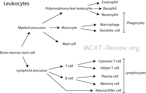
<ul class="fiche-list">- macrophages, neutrophils, mastocytes, cellules tueuses naturelles, cellules dendritiques
- macrophages = phagocytose agent pathogène and then act as cellule présentatrice d'antigène.
- neutrophils = Polymorphonuclear leukocytes = PMNs = phagocytose agent pathogène and destroys it.
- mastocytes: release histamine during an allergic response, bring about inflammation.
- cellules tueuses naturelles: kills infected/abnormal cellules.
- cellules dendritiques: the best cellules présentatrices d'antigène.
- Lymphocytes T
- Matures in the Thymus.
- cellules T cytotoxiques recognize antigène on infected cellules, and signal for apoptose.
- cellules T auxiliaires recognize antigène on cellules présentatrices d'antigène, and signal for activation of cellules B, cellules T, and macrophages.
- Lymphocytes B, plasmocytes
- Matures in Bone marrow.
- cellules B form plasmocytes and cellules mémoire when exposed to antigène.
- plasmocytes = secrete anticorps.
- cellules mémoire = stick around in case the same antigène attacks in the future.
- Tissus
- moelle osseuse
- all cellules sanguines arise from cellules souches in the moelle osseuse.
- lymphocytes B differentiate in the moelle osseuse.
- rate
- Provides a site for WBCs to reside and proliferate.
- Removes agents pathogènes from blood.
- Removes old RBCs and platelets.
- thymus: lymphocytes T differentiate in the thymus.
- ganglions lymphatiques
- Provide a site for WBCs to reside and proliferate.
- Removes agents pathogènes from lymph.
- Residing lymphocytes monitor lymph for foreign antigènes, and initiate an réponse immunitaire when exposed to foreign antigènes.
- Basic aspects of immunité innée and réponse inflammatoire: Innate = first line of defense = kills anything that doesn't look right = not specific to a particular agent pathogène / antigène
- Skin: natural flora, layer of keratin.
- Mucus membranes: traps agent pathogène in mucus, and cilia moves it out.
- Phagocytes: engulf agent pathogène.
- Natural killer cellules: destroy infected cellules.
- Antimicrobial protéines: tears (lyse bacteria), interferons (interfere with virus réplication), complement (punches holes in cellule/agent pathogène membrane).
- Fever/inflammation: WBCs are more active at higher temperature, and inflammation recruits WBCs to site of infection by sending out chemical signals and making capillaires more permeable.
- Adaptive immunité = highly specific for a particular agent pathogène / antigène.
- cellules présentatrices d'antigène present foreign antigène on their surface.
- antigène is recognized by T and cellules B.
- cellules T cytotoxiques kill infected cellules.
- cellules T auxiliaires activate macrophages, T and cellules B.
- cellules B produce anticorps.
- anticorps bind to antigènes and bring about
- neutralization: agent pathogène can't adhere to cellule hôte
- opsonization: makes it easier for phagocytose
- complement activation: kills infected cellule by punching holes in membrane cellulaire.
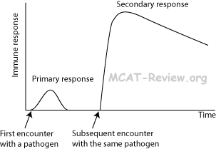
- cellules mémoire are made that are much more efficient (does not need cellule T activation) in proliferating and making anticorps in case the same infection strikes in the future.
- cellules mémoire allow the body to mount a greater, and more sustained response against the same agent pathogène during secondary response.
- Concept of antigène and anticorps
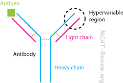
- Anticorps = lock, Antigène = key. Each anticorps is specific to the binding of an antigène.
- Anticorps is like a Y, the tips of the fork bind antigène.
- The tips of the fork are called hypervariable regions because they are unique to each antigène-specific anticorps.
- The anticorps consists of 2 light chains and 2 heavy chains linked together by ponts disulfure.
- Structure of anticorps molecule
- Mechanism of stimulation by antigène; antigène presentation
- agent pathogène enters cellule présentatrice d'antigène (APC)
- pieces of the agent pathogène gets displayed at the surface of APCs.
- cellule T récepteurs recognize the presented antigène, and activates various réponse immunitaires.
- scenario 1: extracellular agent pathogène
- macrophage engulfs agent pathogène.
- pieces of the agent pathogène becomes the antigène and gets presented at the macrophage's surface cellulaire.
- cellules T auxiliaires recognize the presented antigène, and activates macrophages to destroy agent pathogène. Helper cellules T also activate cellules B to produce anticorps against the agent pathogène.
- scenario 2: intracellular agent pathogène
- agent pathogène invades cellule hôte.
- pieces of the agent pathogène gets presented on the host surface cellulaire.
- cellules T cytotoxiques recognize the presented antigène, and signals the infected cellule to self-destruct.
      <div class="source-note">Contenu adapté de <a href="https://mcat-review.org/circulatory-lymphatic-immune-systems.php" target="_blank" rel="noopener">mcat-review.org</a> — Traduction et mise en forme par Logopoïos</div>
    </div>
  </div>

  <div class="chapter" id="ch-2">
    <div class="chapter-header" onclick="toggleChapter(this)">
      <div>
        <div class="chapter-num">Chapitre 02</div>
        <div class="chapter-title">Système digestif</div>
      </div>
      <div class="chapter-toggle">▼</div>
    </div>
    <div class="chapter-body">

      <div class="fiche-section">
        <div class="fiche-section-title">Ingestion</div>
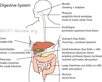
<ul class="fiche-list">- saliva dissolves food.
- saliva contains mucin, a protéine that lubricates the bolus (chewed up food ball).
- saliva contains amylase, which breaks down polysaccharides (starch and glycogen).
- saliva also contains anticorps and lysozyme that kill agents pathogènes.
- epiglottal action
- épiglotte = flap of cartilage that closes off airway when you're swallowing.
- pharynx (function in swallowing)
- pharynx = throat = between mouth and œsophage.
- muscular tube that squeezes and routes food to the œsophage when swallowing (closes off pathways to nasal cavity and airway).
- œsophage (transport function)
- muscular tube that propels bolus (food) to the estomac by péristaltisme.
- péristaltisme = squeezing stuff through a tube (œsophage/gut) by muscle lisse.
      <div class="fiche-section">
        <div class="fiche-section-title">Estomac</div>
<ul class="fiche-list">- storage = the estomac is a muscular bag that is elastic and can stretch to store food.
- churning = mechanical digestion = mixing food.
- low pH, gastric juice, protection by mucus against self-destruction
- Parietal cellules secrete HCl that causes the pH to be very acidic.
- Gastric juice = HCl + pepsin + hormones = secreted by the estomac (parietal and chief cellules, and enteroendocrine cellules)
- Pepsin = protease that works best in acidic environment.
- Goblet cellules secrete mucus lining that protect the estomac from the acid and self-digestion.
- production of enzymes digestives, site of digestion
- Chemical digestion: Estomac produces pepsin, which digests protéines (secreted in an inactive form, gets activated in acidic environment)
- Pepsin is special in that it works best at very acid pH.
- Mechanical digestion: Estomac churns food.
- structure (gross)
- banana shaped bag that can stretch.
- inner membrane densely folded (rugae), so can accommodate stretching.
- sealed off on the top by the cardiac (gastroesophageal) sphinctor.
- sealed off on the bottom by the pyloric sphinctor.
      </div>

      <div class="fiche-section">
        <div class="fiche-section-title">Foie</div>
<ul class="fiche-list">- role in nutrient métabolisme, vitamin storage
- Makes and stores glycogen from glucose.
- Néoglucogenèse from glycerol and acides aminés (deamination).
- Breaks down fats, makes cholesterol, makes lipoproteins used to transport fats.
- Stores vitamins (A, D and B12) and iron.
- Detox: metabolize alcohol, remove ammonia in blood.
- role in glycémie régulation, détoxification
- Blood glucose régulation by foie:
- Blood sugar too low: néoglucogenèse.
- Blood sugar too high: glycogeneis.
- Détoxification: metabolize alcohol (alcohol dehydrogenase), remove blood ammonia, inactivate various other drugs/toxins.
- structure (gross): largest gland in body, spans both sides of the abdomen (though right side much larger). Ducts draining to duodenum and vésicule biliaire.
      </div>

      <div class="fiche-section">
        <div class="fiche-section-title">Bile</div>
<ul class="fiche-list">- Gall vessie stores excess, unused bile, and concentrates it. Secretes it when needed.
- function: bile is an emulsifying agent (not an enzyme). Bile breaks down large fat droplets into smaller microscopic droplets by forming micelles. This increases the total surface of the fat for lipase action.
      </div>

      <div class="fiche-section">
        <div class="fiche-section-title">Pancréas</div>
<ul class="fiche-list">- Pancréas is the major source for all the enzymes digestives.
- Amylase - digests starch.
- Various proteases.
- Lipase - digests fat.
- Ribonuclease - digests acides nucléiques.
- Pancréas makes HCO3- to neutralize the HCl from the estomac.
- transport of enzymes to intestin grêle
- Digestive enzymes of pancréas = exocrine = flows into intestin grêle via duct.
- structure (gross): tadpole-shaped gland with duct leading to duodenum.
      </div>

      <div class="fiche-section">
        <div class="fiche-section-title">Intestin grêle</div>
<ul class="fiche-list">- Small intestin is the major place for digestion and absorption.
- Folds, villi, and microvilli increases the surface for absorption.
- Absorbs digested food into circulation (fats into lacteals, all others into capillaires).
- Active transport occurs to absorb against the gradient de concentration.
- Intestinal lumen (less glucose) -> enterocyte (more glucose): Secondary transport actif by Na+-K+ pump + Na+-Glucose symport.
- Passive/diffusion facilitée occurs to absorb down the gradient de concentration.
- Enterocyte (more glucose) -> liquide extracellulaire (less glucose): Facilitated diffusion (then the glucose will go from the liquide extracellulaire to blood).
- function and structure of villi
- Villi = finger-like protrusions inside intestin grêle.
- Microvilli = same as villi but on the surface of a single absorptive cellule.
- production of enzymes, site of digestion
- The intestin grêle is the major place for digestion and absorption.
- Pancréas is the major source for enzymes. However, the intestin grêle does make some of its own enzymes, including protease and amylase.
- neutralization of acide gastrique
- The pancréas makes bicarbonate ion to neutralize the HCl from the estomac.
- This neutralization facilitates enzymes in the intestin grêle, which would be denatured by estomac pH.
- structure (anatomic subdivisions)
- Duodenum.
- Jejunum.
- Ileum.
      </div>

      <div class="fiche-section">
        <div class="fiche-section-title">Gros intestin</div>
<ul class="fiche-list">- Cecum: blind pocket containing appendix.
- Ascending colon
- Transverse colon
- Descending colon
- Sigmoid colon
- Rectum: stores feces.
- absorption of water: The gros intestin absorbs any remaining water that is not absorbed by intestin grêle.
- bacterial flora
- Ferment undigested nutriments, make gas.
- Produce vitamin K (important for clotting).
- structure (gross): lobes/pockets along its length due to muscle tone. Unlike intestin grêle, the gros intestin has no folds or villi.
      </div>

      <div class="fiche-section">
        <div class="fiche-section-title">Rectum (stockage et élimination des déchets, fèces)</div>
<ul class="fiche-list"> </div>
<div class="fiche-section">
<div class="fiche-section-title">Contrôle musculaire</div>
<ul class="fiche-list">- Cardiac sphincter (gastroesophageal sphincter): sphincter between œsophage and estomac. Prevents back flow of food.
- Pyloric sphincter: between estomac and intestin grêle. Releases food into the intestin grêle a small amount at a time.
- Anal sphincter: at the end of rectum. ties the end of the rectum.
- péristaltisme: involuntary movement of muscles lisses, squeezes food along the tube digestif.
      </div>

      <div class="source-note">Contenu adapté de <a href="https://mcat-review.org/digestive-system.php" target="_blank" rel="noopener">mcat-review.org</a> — Traduction et mise en forme par Logopoïos</div>
    </div>
  </div>

  <div class="chapter" id="ch-3">
    <div class="chapter-header" onclick="toggleChapter(this)">
      <div>
        <div class="chapter-num">Chapitre 03</div>
        <div class="chapter-title">Biologie moléculaire : Enzymes et métabolisme</div>
      </div>
      <div class="chapter-toggle">▼</div>
    </div>
    <div class="chapter-body">

      <div class="fiche-section">
        <div class="fiche-section-title">Structure et fonction des enzymes</div>
<ul class="fiche-list">- Enzymes are catalyseurs, which are things that increase the rate of a reaction, but does not get used up during the reaction.
- Structure determines function. A change in structure => a change in function.
- Important biological reactions catalyzed by enzymes:
- Métabolisme
- synthèse d'ADN
- RNA synthesis
- Protéine synthesis
- Digestion
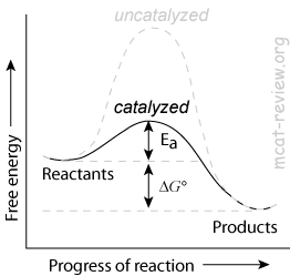
- Reduction of énergie d'activation
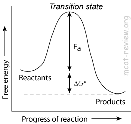
- Enzymes decrease the énergie d'activation (Ea) of a reaction by lowering the energy of the transition state.
- Enzymes increase the rate of a reaction by decreasing the énergie d'activation.
- Enzymes will increase the rate constant, k, for the equation rate = k[A][B].
- Enzymes do NOT change the Keq of a reaction.
- Enzymes do not change Keq because it lowers the énergie d'activation for BOTH forward and reverse reactions.
- Enzymes will make the reverse reaction go faster also.
- Enzymes do not change ΔG, the net change in free energy.
- Enzymes affect the kinetics of a reaction, but not the thermodynamics.
- Substrats and enzyme specificity
- Enzyme-substrat interactions occur at the enzyme's active site.
- Enzyme-substrat specificity derives from structural interactions.
- Lock and key model: rigid active site. Substrat fits inside the rigid active site like a key.
- Induced fit model: flexible active site. Substrat fits inside the flexible active site, which is then induced to "grasp" the substrat in a better fit.
- Enzymes can be specific enough to distinguish between stereoisomers.
- Enzymes can be protéine or RNA.
- Almost all enzymes in your body is made of protéine.
- The most important RNA enzyme in your body is the ribosome.
- Enzyme structure derives from 4 levels.
- Primary: this is the sequence of the protéine or RNA chain.
- Secondary: this is liaison hydrogène between the protéine backbone. Examples include alpha helices and beta sheets (backbone H-bonding). For RNA, this is paire de basesing.
- Tertiary: this is the 3-D structure of the enzyme. This involves -R group interactions and spatial arrangement of secondary structure.
- Quaternary: when more than 1 chain is involved. When you hear about "dimers", "trimers", "tetramers", "oligomers", that's quaternary structure.
- Heat and extreme pH denatures enzymes by altering their structure.
<div class="fiche-section">
<div class="fiche-section-title">Contrôle de l'activité enzymatique</div>
<ul class="fiche-list">- The produit of a pathway inhibits the pathway.
- For example, hexokinase, the first enzyme in glycolyse, is inhibited by its produit glucose-6-phosphate.
- Competitive inhibition
- An inhibiteur competes with the substrat for binding to the active site.
- Competitive inhibition increases the amount of substrat needed to achieve maximum rate of catalysis.
- Competitive inhibition does NOT change the maximum possible rate of the enzyme's catalysis.
- You can overcome competitive inhibition by providing more substrat.
- Non-competitive inhibition
- An inhibiteur binds to an allosteric site on the enzyme to deactivate it.
- The substrat still have access the active site, but the enzyme is no longer able to catalyze the reaction as long as the inhibiteur remains bound.
- Non-competitive inhibition decreases the maximum possible rate of the enzyme's catalysis.
- Non-competitive inhibition does NOT change the amount of substrat needed to achieve the maximum rate of catalysis.
- You can't overcome non-competitive inhibition by adding more substrat.
      <div class="fiche-section">
        <div class="fiche-section-title">Métabolisme de base</div>
<ul class="fiche-list">- Steps of métabolisme aérobie (needs oxygen)
- Glycolyse
- Oxidative decarboxylation
- Krebs cycle
- Electron transport chain.
- Steps of métabolisme anaérobie (don't need oxygen)
- Glycolyse
- Alcohol or acide lactique fermentation
- Aerobic métabolisme of glucose
- Complete oxidation of metabolite (glucose) to dioxyde de carbone.
- ~30 ATP produced per glucose.
- C6H12O6 + 6O2 => 6CO2 + 6H2O
- C6H12O6: this is glucose. You get it from your diet.
- 6O2: this is oxygène moléculaire that you breathe in.
- 6CO2: this is dioxyde de carbone produced by the Krebs cycle. Both the carbon and oxygen in this CO2 comes from the metabolite (glucose).
- 6H2O: this is water produced in the chaîne de transport d'électrons. The oxygen comes completely from the oxygène moléculaire that you breathe in.
- If we were to follow the carbon in the metabolite (glucose), it will end up in dioxyde de carbone.
- If we were to follow the oxygen in the metabolite (glucose), it will end up in dioxyde de carbone.
- If we were to follow the oxygen you breathe in, it will end up in water.
- As for the hydrogens, they'll either be in water, exist as protons in solution, or be transferred to some other entity.
- As we can see, the total reaction involves complete oxidation of the metabolite (glucose) and complete reduction of oxygène moléculaire.
- When electrons pass from the metabolite (glucose) to oxygène moléculaire, energy is released.
- The chaîne de transport d'électrons harnesses this energy.
- Anmétabolisme aérobie of glucose
- Partial oxidation of metabolite (glucose) to pyruvate.
- 2 net ATP produced per glucose.
- Pyruvate is then reduced to either alcohol or lactate.
- Bacteria reduce pyruvate to alcohol in a process called alcohol fermentation.
- Humans reduce pyruvate to lactate in a process called acide lactique fermentation.
- Glycolyse, anaerobic and aerobic, substrats and produits
- Glycolyse = convert glucose (6 carbons) to 2 molecules of pyruvate (3 carbons).
- Location: cytosol.
- 2 net ATP made for every glucose (2 input ATP, 4 output ATP).
- 2 NADH made for every glucose.
- Occurs under both aerobic and conditions anaérobies.
- Glycolyse is inhibited by ATP.
- Aerobic decarboxylation (mitochondrial matrix) = convert pyruvate (3 carbons) to an acetyl group (2 carbons).
- 1 NADH made for every pyruvate.
- Only occurs in the presence of oxygen.
- Acetyl group attaches to Coenzyme A to make acetyl CoA.
- Anaerobic fermentation (cytosol) = redox reaction: reduce pyruvate, oxidize NADH.
- 1 NAD+ made for every pyruvate.
- Alcohol fermentation = pyruvate reduced to ethanol.
- Lactic acid fermentation = pyruvate reduced to lactate.
- The purpose of anaerobic fermentation is to regenerate NAD+, which is needed for glycolyse.
- Krebs cycle, substrats and produits, general features of the pathway
- Location: matrix of mitochondries.
- Acetyl CoA feeds into the cycle.
- 3 NADH made per acetyl CoA.
- 1 FADH2 made per acetyl CoA.
- 1 ATP (GTP) made per acetyl CoA.
- Coenzyme A is regenerated (during the first step of the cycle).
- Krebs cycle, TCA, Tricarboxylic acid cycle, citric acid cycle all mean the same thing.
- Krebs cycle is Inhibited by ATP and NADH.
- Electron transport chain and phosphorylation oxydative, substrats and produits, general features of the pathway
- Location: the cristae (inner membrane of mitochondries).
- Input NADH
- Proton gradient
- The chaîne de transport d'électrons (ETC) is essentially a series of redox reactions, where NADH gets oxidized to NAD+ and O2 gets reduced to H2O.
- The series of redox reactions consists of electrons passing from NADH to FMN, to Coenzyme Q, iron-sulfur complexes, and cytochromes (cytochrome b, c and aa3) before finally being used to reduce oxygen.
- NADH is highest in energy, while O2 is lowest in energy. When electrons are passed from NADH down a series of protéines and finally to O2, energy is released.
- FADH2 is lower in energy than NADH, that's why it releases less energy when it gets oxidized.
- FADH2 skips FMN and passes its electrons to Coenzyme Q.
- The energy released from these reactions generates a gradient de protons, which drives ATP synthase to make ATP. This is called phosphorylation oxydative.
- Proton gradient
- The energy released from passing electrons down the ETC is used to pump protons into the intermembrane space of the mitochondries.
- H+ concentration is very high in the intermembrane space (higher than those in the matrix). Thus, this establishes an gradient électrochimique called the gradient de protons.
- H+ wants to migrate down the gradient de protons (from the intermembrane space back into the matrix), but it can only do this by going through the ATP synthase.
- Like a water mill, ATP synthase harnesses the energy of the falling protons to convert ADP into ATP.
- The ETC is inhibited by certain antibiotics, by cyanide, azide, and monoxyde de carbone.
- Métabolisme of fats and protéines
- Fat métabolisme
- Location: beta-oxidation occurs in the matrix of the mitochondries. Ester hydrolysis occurs in the cytosol.
- Fatty esters gets hydrolyzed into free acides gras by lipases.
- For example, triacylglycerol gets hydrolyzed into free acides gras and glycerol.
- With the help of ATP, the acide gras is "activated" at the acid end by CoA (to be precise, it turns into a thioester).
- A process called beta-oxidation breaks down the fatty-CoA, 2 carbons at a time, to make acetyl CoA.
- β-oxidation produces acetyl CoA and also FADH2 and NADH.
- The acetyl CoA feeds into the Krebs cycle, and the FADH2 and NADH feed into the ETC.
- On a per gram basis, fats give more energy than any other food source.
- Protéine métabolisme
- Protéines are broken down into acides aminés by peptidases.
- The nitrogen in the acide aminé is converted to urea (for desert animals, birds and reptiles, it is acide urique).
- The carbon in the acide aminé is converted to pyruvate or acetyl-CoA, (or other metabolical intermediates such as oxaloacetate), depending on what acide aminé it is.
- The carbon produits from acide aminé métabolisme can either feed into the Krebs cycle, or be the starting material for néoglucogenèse.
      </div>

      <div class="source-note">Contenu adapté de <a href="https://mcat-review.org/enzymes-metabolism.php" target="_blank" rel="noopener">mcat-review.org</a> — Traduction et mise en forme par Logopoïos</div>
    </div>
  </div>

  <div class="chapter" id="ch-4">
    <div class="chapter-header" onclick="toggleChapter(this)">
      <div>
        <div class="chapter-num">Chapitre 04</div>
        <div class="chapter-title">Évolution</div>
      </div>
      <div class="chapter-toggle">▼</div>
    </div>
    <div class="chapter-body">

      <div class="fiche-section">
        <div class="fiche-section-title">Évolution</div>
<ul class="fiche-list">- Fitness concept
- Fitness is defined as the ability to pass your gènes on, or reproductive success.
- The classical trick question gives you an individual who is strong, healthy, long-living, but does not reproduce. In this case, no matter how good the other traits are, if the individual does not reproduce, then it has a fitness of zero.
- Selection by differential reproduction
- Individuals who reproduce more viable offspring are selected for.
- Individuals who reproduce less viable offspring are selected against.
- Concepts of natural and group selection
- Natural selection = survival and reproduction of the fittest.
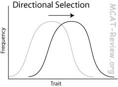
Directional selection: selects for a trait on one extreme. For example, selection for height of canopy trees in a rainforest: trees compete for sunlight, so selection favors trees to become higher and higher.
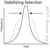
Stabilizing selection: selects for a trait that is moderate, and selects against the extremes. For example, birthweight: too low birthweight means that the baby is premature, too high birthweight means that the mom will have a hard time delivering, so there's a "just right" birthweight that is selected for.
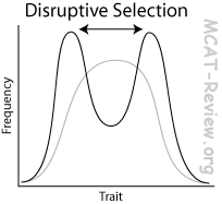
Disruptive selection: selects for the extremes. For example, birds occupying a habitat with 2 distinct niches (eating berries for a living and eating seeds for a living): small beaks are selected for eating berries, large beaks are selected for cracking seeds, medium beak is left out.
- Group selection = sélection naturelle acting on the group, not the individual.
- Explains why altruisme exists.
- Altruisme sacrifice the fitness of the individual to benefit the group (family), which shares similar gènes with the individual. When the benefit outweighs the cost, the altruistic behavior is selected for.
- Evolutionary success as increase in percent representation in the pool génique of the next generation
- If the frequency of an allèle increased, then that's evolutionary success for that allèle.
- If the frequency of allèles of an individual increased in a population, then that's evolutionary success for that individual.
- Spéciation
- Definition of species
- Three conditions for biological species
- Be able to interbreed.
- Be able to produce fertile, viable offspring.
- Does this naturally.
- A dog and a cat can't interbreed, so they don't belong to the same species.
- A horse and a donkey can interbreed, but their offspring, the mule, is sterile. So horses and donkeys aren't the same species.
- Some species of flowers can cross pollinate to produce fertile offspring. However, this never occurs in nature because one is bee-pollinated and the other is bird-pollinated. Thus, they are different species even though they can potentially produce fertile offspring.
- Spéciation is the formation of a new species. This can occur due to barriers to successful interbreeding within an initial species.
- Polymorphisme
- Polymorphisme is just a fancy word for different forms of allèles/traits.
- Adaptation and spécialisation
- Adaptation is the genetic change in a population caused by sélection naturelle.
- Adaptation is caused by Darwin's sélection naturelle, not by Lamarck's ideas. A giraffe's neck is long because long necks increase the survival rate, so more long-necked giraffes survive to reproduce, and over many generations, the population evolved long necks. The wrong idea by Lamarck is that the giraffe had to reach for higher leaves on trees, so it stretched itself a longer neck.
- Spécialisation = adaptation of traits to better fill a niche.
- Concepts of ecological niche, competition
- A species' ecological niche is what resources the species uses to survive in its environment.
- Two species can avoid competition, and better use the environment's resources by occupying different niches.
- As long as two species occupy different niches, there's no competition because they use different resources.
- When niches overlap, there's competition.
- Spécialisation occurs to better occupy a particular niche.
- Concept of population growth through competition
- Population growth is checked by competition.
- When resources get scarce, competition increases, which slows down population growth.
- Competition within a species can force members within the species to occupy different niches, which drives spéciation.
- Consanguinité
- Consanguinité is mating between relatives.
- Consanguinité increases the frequency of homozygotes, decreases heterozygotes, and decreases diversité génétique.
- Consanguinité depression occurs because of the increase in the frequency of homozygous recessive detrimental allèles.
- Some species (naked mole rats) naturally inbreed because:
- They stay in one small area and don't migrate much.
- Detrimental homozygous allèle récessifs are eliminated because of many generations of sélection naturelle.
- Exogamie
- Exogamie is mating with non-relatives, which is just the opposite of consanguinité.
- Exogamie increases heterozygosity.
- Goulots d'étranglement, dérive génétique
- A goulot d'étranglement is a severe reduction in population size. This can be caused, for example, by a natural disaster that wipes out a majority of the population.
- Genetic drift is the random changes in fréquences alléliques.
- The effect of dérive génétique increases as population size decreases.
- Goulots d'étranglement increase the effect of dérive génétique.
- Divergent, parallel, and convergent évolution
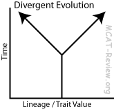
Divergent évolution
- Same lineage, evolving apart to be more different.
- For example, bats and horses. Both share the same lineage as mammals, but the limb of the bat became wings while the horse developed hooves.
- Divergent évolution produces homologous structures (bat's wing and horse's hoof).
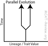
Parallel évolution
- Same lineage, evolving closer together to be similar, using similar mechanisms.
- For example, the feeding structure in different species of crustaceans. The feeding structure came from mutation of pair of legs, turning them into mouth parts. This is a prime example of parallel évolution: same lineage, similar traits, evolved from similar mechanisms/mutations.
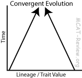
Convergent évolution
- Different lineage, evolving closer together to be similar, using different mechanisms.
- For example, bats and butterflies. Both have wings, but they came from totally different lineages, evolved through different mechanisms/mutations. Convergent évolution produces analogous structures (bat's wing and butterfly's wing).
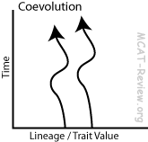
Coevolution
- Two species evolve in response to each other.
- For example, predator/prey or host/parasite species.
- Not yet an official MCAT topic, but many students confuse parallel évolution with coevolution.
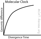
- Evolutionary time as measured by gradual random changes in genome
- Random genetic mutations (drift) that are not acted on by sélection naturelle (neutral) occur at a constant rate.
- By measuring the amount of these neutral mutations, you can find out how much time has passed.
- You can compare genome differences between two species to find out how long ago they diverged.
- Another name for this concept is the Molecular Clock.
      <div class="fiche-section">
        <div class="fiche-section-title">Évolution</div>
<ul class="fiche-list">- Parasitism
- Relationship where one benefits (parasite), and the other is harmed (host).
- For example, worms living inside animal intestines.
- Commensalism
- Relationship where one benefits, and the other is not affected.
- For example, some plant seeds disperse by sticking to animal fur.
- Mutualism
- Relationship where both species benefit.
- For example, lichens are made from a mutualistic relationship between fungi and algae. The fungus provides anchor/absorption, and the alga provides photosynthèse.
- Relationship between ontogeny and phylogeny
- Ontogeny = development through the life of an organism.
- Phylogeny = development through evolutionary time of lineages/species.
- In early development, vertebrate embryos share similar features, reminiscent of a common ancestor.
- Gill slits
- Notochord
- Segmentation
- Paddle-like limbs
- Ontogeny recapitulates phylogeny is the idea that the development of an organism repeats the evolutionary history of its species; starting with the fish-like common ancestor, which then changes to the modern form as development continues to adulthood.
- Origin of life
- Organic molecules created by atmospheric gases zapped by lightning, which falls into the ocean to make primordial soup (Oparin and Haldane). Urey-Miller's experiment proved this in a lab.
- RNA World hypothesis: the simple organic molecules formed RNA polymers that can self-replicate (Having enzymatic activity as well as serving as template).
- Protocells: aggregates of RNA, protéines inside lipid envelopes.
- Prokaryotes: first anaerobic heterotrophs because early atmosphere blocks the light required for photosynthèse, then anaerobic autotrophs that undergoes photosynthèse and makes oxygen, then aerobics that utilize oxygen.
- Eukaryotes: evolved by endosymbiose, where a big cellule engulfed a smaller cellule and then developed a mutualistic relationship. Heterotrophs engulfed mitochondries. Autotrophs engulfed chloroplasts.
      <div class="fiche-section">
        <div class="fiche-section-title">Anatomie comparée</div>
<ul class="fiche-list">- Chordate features
- Notochord = the "backbone" of the embryon, except that it's not made of bone. In vertebrates, bones will replace the notochord to form the vertebrae.
- Pharyngeal pouches, branchial arches (pharangeal pouches) = gill slits in the embryon. Later develop into various head and neck structures in human.
- Dorsal nerf cord = forms the système nerveux. In higher chordates, the nerf cord develops into the cerveau and moelle épinière.
- Vertebrate = a group of chordates (subphylum).
- Vertebrate phylogeny: vertebrate classes and relations to each other
- Fish: In the beginning, there was fish.
- Jawless (Agnatha): The very first fish were jawless, slimy, eel-like.
- Cartilaginous (Condrichthyes/sharks/rays): Then some developed jaws and a skeleton. Condrichthyes has a skeleton made of cartilage.
- Bony (Osteichthyes/food fish): Osteichthyes has a skeleton made of bone.
- Amphibians: the Bony Fish came onto land because their bony skeleton is strong enough to support their weight.
- Reptiles: Can penetrate further onto land because they don't dry out like amphibians do. Similar to amphibians, the reptiles lay eggs.
- Mammals: First to branch off from the reptiles. Unlike the reptiles, mammals have milk glands, hair, and different tooth morphology (heterodontic).
- Birds: next to branch off from the reptiles. Like the reptiles, birds lay eggs. (which came first, the chicken or the ovule? Ans: the ovule, because the chicken is a bird, and reptiles laid eggs before birds even existed.)
<div class="source-note">Contenu adapté de <a href="https://mcat-review.org/evolution.php" target="_blank" rel="noopener">mcat-review.org</a> — Traduction et mise en forme par Logopoïos</div>
</div>
</div>
<div class="chapter" id="ch-5">
<div class="chapter-header" onclick="toggleChapter(this)">
<div>
<div class="chapter-num">Chapitre 05</div>
<div class="chapter-title">Système excréteur</div>
</div>
<div class="chapter-toggle">▼</div>
</div>
<div class="chapter-body">
<div class="fiche-section">
<div class="fiche-section-title">Rôles dans l'homéostasie</div>
<ul class="fiche-list">- If pression artérielle too low:
- Renin-angiotensin pathway: Rein (JGA cellules) release renin, triggers formation of angiotensin II, which stimulates aldosterone release, the end result is to raise pression artérielle.
- Aldosterone (aka mineralocorticoid): Adrenal glands release aldosterone, causes rein (tubules distaux) reabsorb more Na+, which in turn causes more réabsorption d'eau.
- ADH (made in hypothalamus, stored in pituitary): causes more réabsorption d'eau in the rein tubules, raising pression artérielle. High levels also cause vasoconstriction.
- If pression artérielle too high, all the above hormones stop releasing. Also, the heart can release ANP (Atrial natriuretic peptide), which antagonizes aldosterone and cause rein to excrete both more Na+ and more water. ANP can also cause vasodilatation.
- osmorégulation
- Blood plasma is mainly Na+ and Cl- (Inside cellules is mainly K+ and hydrogen phosphate ions).
- Blood osmolarity is determined predominantly by Na+ and Cl-.
- Blood osmolarity too low → aldosterone, reabsorb Na+. Cl- follows.
- Rein tubules' sécrétion and reabsorption regulates osmolarity.
- Other ions
- K+ is regulated by aldosterone.
- Aldosterone: reabsorb Na+, pee out K+
- Calcium and phosphate regulated by PTH.
- PTH = parathyroid hormone = more Ca2+ reabsorption in rein tubules (also, bone break down to release calcium and phosphates, and intestin grêle to absorb more calcium).
- acid-base balance: keep pH sanguin constant.
- Buffer systems: Bicarbonate buffer system (blood and liquide extracellulaire), Phosphate Buffer System (inside cellules)
- CO2 + H2O ⇌ H2CO3 ⇌ H+ + HCO3-
- Breathing out CO2 decreases the acidity in blood.
- Rein tubules:
- Bicarbonate ion (HCO3-): peeing it out makes blood more acidic. Reabsorption makes blood more basic. (not the other way round, use Le Chatelier's principle, or simply note that bicarbs bind H+, so they have the opposite effect on pH)
- H+ sécrétion gets rid of acidity.
- removal of soluble nitrogenous waste
- Urine = concentrated urea in water, with some salt.
- Urea = harmless form of toxic ammonia = nitrogenous waste.
- Amino acids → Ammonia → Urea → peed out.
      <div class="fiche-section">
        <div class="fiche-section-title">Structure du rein</div>
<ul class="fiche-list">- medulla = inner part of rein = contains anse de Henlé.
      <div class="fiche-section">
        <div class="fiche-section-title">Structure du néphron</div>
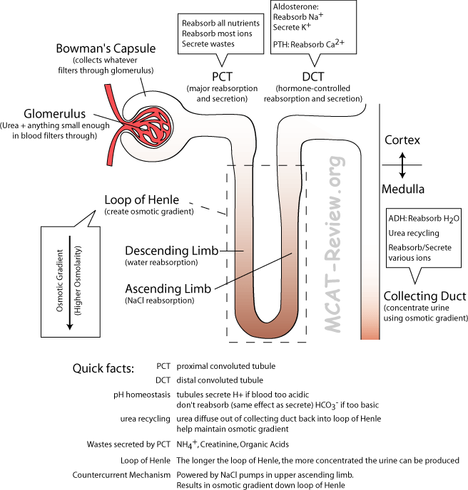
<ul class="fiche-list">- glomérule = ball of fenestrated capillaires.
- capsule de Bowman = Cup/Capsule that surrounds the glomérule.
- tubule proximal = tubule contourné on the side of the capsule de Bowman = the major site for reabsorption (nutrient, salts and water) and sécrétion (except for K+, the sécrétion of which is the job of tubule distal contourné in response to aldosterone).
- anse de Henlé = U shaped loop that dips into the médulla rénale = multiplicateur à contre-courant mechanism occurs here
- Descending limb = réabsorption d'eau by osmose (permeable to water, but not to solute).
- Bottom of U = most concentrated.
- Ascending limb = réabsorption de sel (permeable to salt, but not water).
- tubule distal = tubule contourné on the side of the canal collecteur = hormone-controlled (fine tunes the work done by the tubule proximal) reabsorption of salts and water. Aldosterone-controlled sécrétion of K+
- canal collecteur = the tubules distaux of many nephrons drain here = ADH-controlled reabsorption of water, hormone-controlled reabsorption/sécrétion of salts.
</div>
<div class="fiche-section">
<div class="fiche-section-title">Formation de l'urine</div>
<ul class="fiche-list">- Powered by pression hydrostatique.
- Both good stuff, bad stuff and ions are filtered out, as long as it's small enough.
- Good stuff: nutriments
- Bad stuff: urea (and creatinine and acide urique)
- Good stuff reabsorbed, bad stuff peed out.
- sécrétion and reabsorption of solutes
- Proximal tubules contournés reabsorb all the good stuff (nutriments) and most of the ions. Bad stuff left in the filtrate (urea) to be peed out, also actively excreted (NH4+, creatinine, organic acids).
- Loop of Henle reabsorbs water and salt using the mécanisme à contre-courant.
- Distal tubules contournés selectively reabsorb or secrete stuff based on hormonal control.
- Collecting duct reabsorb water to concentrate urine if ADH present. (Also can secrete and reabsorb stuff based on hormonal control)
- Régulation of pH sanguin: secrete H+ when blood too acidic, pee out (don't reabsorb) HCO3- when blood too basic.
- concentration of urine
- The tubule distal contourné contains dilute solution of urea.
- The canal collecteur concentrates it by réabsorption d'eau (diffusion facilitée) when ADH is present.
- Water reabsorption in the canal collecteur is possible because the anse de Henlé has very high osmolarity (very concentrated) at the bottom.
- multiplicateur à contre-courant mechanism (basic function)
- What does the Countercurrent multiplier do? It creates an gradient osmotique down the anse de Henlé, which is used by the canal collecteur to concentrate urine.
- What drives the creation of this gradient? NaCl pump on ascending limb.
- What's countercurrent? Descending limb: water flow out of filtrate, impermeable to salt. Ascending limb: salt flow out of filtrate, impermeable to water.
- What's multiplier? The gradient-producing power of each individual NaCl pump multiplies down the length of the anse de Henlé. Longer the anse de Henlé, greater the gradient osmotique, more concentrated urine can be produced.
- What is urea recycling? Urea at the bottom of canal collecteur leaks out into the liquide interstitiel and back into the filtrate. Contributes to the high osmolarity at the bottom of the anse de Henlé.
      </div>

      <div class="fiche-section">
        <div class="fiche-section-title">Stockage et élimination (uretère, vessie, urètre)</div>
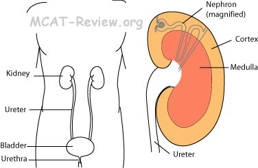
<ul class="fiche-list">- Uretères drain into the vessie.
- Vessie stores urine: its special epithelium (transitional epithelium) can squish to accommodate storage of large amounts of urine.
- Urine gets peed out of the vessie through the urètre.
      </div>

      <div class="source-note">Contenu adapté de <a href="https://mcat-review.org/excretory-system.php" target="_blank" rel="noopener">mcat-review.org</a> — Traduction et mise en forme par Logopoïos</div>
    </div>
  </div>

  <div class="chapter" id="ch-6">
    <div class="chapter-header" onclick="toggleChapter(this)">
      <div>
        <div class="chapter-num">Chapitre 06</div>
        <div class="chapter-title">Cellule eucaryote généralisée</div>
      </div>
      <div class="chapter-toggle">▼</div>
    </div>
    <div class="chapter-body">

      <div class="fiche-section">
        <div class="fiche-section-title">Membrane plasmique</div>
<ul class="fiche-list">- Composition of membranes
- Lipid components
- Phospholipids (forms bilayer)
- Steroids, cholesterol (provide more fluidity)
- Waxes (provide more stability)
- Protéine components: récepteurs, channels, transport protéines
- Fluid mosaic model: the fluid mosaic model basically describes the membrane as protéine boats floating in a sea of lipids.
- Membrane dynamics
- Invaginates to take things in: phagocytose, pinocytose, endocytose
- Buds off: exocytose
- Changes shape: chemotaxis with the aid of changes in cytoskeleton
- Let's things through: small nonpolar molecules, others by the aid of channels or pumps
- Solute transport across membranes
- Thermodynamic considerations
- Mixing a charged ion with hydrophobilic lipid bilayer is thermodynamically unfavorable by entropy
- Therefore, ions can't pass through the lipid bilayer without assistance from channels/pumps
- Osmose: water diffuses freely across the membrane, but not ions. So osmose occurs readily
- Colligative properties depend on the number of solute dissolved in solvent
- Solvent will move as to dilute the component with more solute dissolved (given a semipermeable membrane that lets solvent pass but not the solutes)
- Osmotic pressure = pressure exerted by solvent via osmose = counter pressure needed to prevent osmose
- The bigger the difference in solute concentration, the bigger the pression osmotique
- Too much pression osmotique, and the cellule will lyse (eg: if you put animal cellules in water)
- Passive and transport actif: things that can't readily diffuse across the membrane are transported across the membrane either without energy (passive) or with energy (active).
- Passive transport = no ATP needed = channels, diffusion facilitée = direction of movement is down a gradient de concentration
- Active transport = ATP needed = sodium/ptassium pump = up/against a gradient de concentration
- Sodium-potassium pump: 3 sodium (NA+) out, 2 potassium (K+) in. Thus, the cellule maintains a negative potentiel de repos.
- Membrane channels: lets ions through the membrane cellulaire
- Membrane potential: the potentiel de repos of the membrane cellulaire is negative because of the sodium-potassium pump.
- Membrane récepteurs, signalisation cellulaire pathways, seconds messagers
- Many hormones can't cross the membrane plasmique, so they bind to membrane récepteurs on the outside.
- Récepteur binding triggers the production of seconds messagers.
- Second messengers cause a change inside the cellule (through a protéine kinase cascade).
- Cellule signaling pathways:
- Contact signaling = physical contact triggers a change inside cellule.
- Chemical signaling = chemical binding to récepteur triggers a change inside cellule.
- Nerfs use neurotransmetteurs.
- The système endocrinien use hormones.
- Electrical signaling = change in potentiel de membrane triggers change in cellule.
- Action potential along neurones propagates and cause release of neurotransmetteurs into synapse..
- Action potential along muscle membrane cellulaire causes contraction.
- Exocytose and endocytose: exo = getting stuff out, endo = taking stuff in.
- Intercellular junctions
- jonctions communicantes: connects two cellules, and allows stuff to flow through between the cellules.
- jonctions serrées: stitches/glues two cellules together, and does not allow stuff to flow through between the cellules. A series of cellules with jonctions serrées also effectively forms an impermeable barrier.
- desmosomes: connects two cellules together by linking their cytoskeleton. They are organized for mechanical strength, not an impermeable barrier.
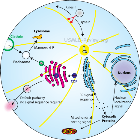
      <div class="fiche-section">
        <div class="fiche-section-title">Organites membranaires et caractéristiques des cellules eucaryotes</div>
<ul class="fiche-list">- Defining characteristics = what sets eukaryotes apart from prokaryotes.
- Eukaryotes have a true noyau (membrane-bound), while prokaryotes don't.
- Eukaryotes have membrane-bound organites (ER, Golgi, lysosomes, mitochondries), prokaryotes don't.
- Eukaryotes divide by mitose (all them chromosomes line up and stuff), prokaryotes undergo fission binaire (no chromosomes, just a circular ring of DNA, no need for complex mitose)
- Noyau (compartmentalization, storage of genetic information)
- compartmentalization: membrane nucléaire / enveloppe nucléaire surrounds the noyau.
- genetic information is stored inside the noyau as DNA.
- Nucléole (location and function)
- location is a region inside the noyau.
- function is to transcribe ribosomal RNA (rRNA).
- Nuclear envelope, pores nucléaires
- enveloppe nucléaire is a double membrane system made of an outer and an inner membrane. Also called membrane nucléaire.
- pores nucléaires are holes in the enveloppe nucléaire where things can pass into and out of the noyau. Transcription occurs in the noyau, and those transcribed RNA need to pass out of the noyau. Things like facteurs de transcription need to pass into the noyau where they can access the DNA to be transcribed.
      <div class="fiche-section">
        <div class="fiche-section-title">Organites membranaires</div>
<ul class="fiche-list">- site of ATP production: an apparatus called the ATP synthase makes ATP from ADP by utilizing the gradient de protons as the driving force. The gradient de protons is where the proton H+ concentration is higher in the inter-membrane space than the matrix of the mitochondries.
- self-réplication; have own DNA and ribosomes.
- mitochondries replicate independently from the cellule containing the mitochondries.
- mitochondries does not share the same genome with its host.
- mitochondries has their own ribosomes, which are different from the host's ribosomes in both sequence and structure.
- All these serve to support the endosymbiose theory.
- inner and outer membrane
- Inner membrane surrounds the matrix.
- The folds of the inner membrane make up the cristae.
- Between the outer and inner membrane is the intermembrane space.
- The intermembrane space is high in protons H+.
- The outer membrane separates the mitochondries from the cytoplasme.
- Lysosomes (vesicle containing hydrolytic enzymes)
- Digests things like food and viral/bacterial particles.
- Things you want to digest gets into a vacuole by endocytose or phagocytose, and then the vacuole fuses with the lysosome. Anything inside gets digested by the hydrolytic enzymes.
- Endoplasmic reticulum:
- rough (RER) and smooth (SER)
- RE rugueux has ribosomes studded over it, RE lisses don't.
- RER deals with synthèse des protéines, folding, modification, and export.
- SER deals with biosynthesis of lipids and steroids, and métabolisme of carbohydrates and drugs.
- In the muscles, the SER or SR stores and regulates calcium.
- RER (site of ribosomes): the ribosomes attach to the outside of RE rugueux and synthesis protéine into the lumen.
- role in membrane biosynthesis: SER (lipids), RER (transmembrane protéines)
- SER = makes lipids of the membrane plasmique.
- RER = makes transmembrane protéines, carries them on its membrane, RER membrane forms vesicles and bud off, fuses with the membrane plasmique, transmembrane protéines now on the membrane plasmique.
- RER (role in biosynthesis of transmembrane and secreted protéines that cotranslationally targeted to RER by séquence signal)
- Transmembrane protéines, or protéines that are to be secreted (need RER vesicle) have a séquence signal right at the beginning.
- When ribosome starts making those protéines, they make the séquence signal first.
- Signal sequence recruits a signal recognition particle that drags it to the RER.
- ribosome now on the RER continues making the protéine, but snakes it into the lumen.
- Signal sequence is clipped off.
- All ERs are connected to the membrane nucléaire (an old aamc topic, no longer tested).
- appareil de Golgi (general structure; role in packaging, sécrétion, and modification of glycoprotein carbohydrates)
- looks like stacks of pancakes.
- modifies and/or secretes macromolecules for the cellule.
- RER make protéine → modified in the Golgi → buds off golgi and secreted out of cellule by exocytose.
- Glycoprotein = protéine with attached saccharides.
- Golgi can glycosylate protéines as well as modifying existing glycosylations.
- Glycosylation affects protéine's structure, function, and protect it from dégradation.
- Peroxisomes: organites that collect peroxides
- Contain oxidative enzymes for intracellular digestion
      </div>

      <div class="fiche-section">
        <div class="fiche-section-title">Cytosquelette</div>
<ul class="fiche-list">- Microfilaments (composition; role in cleavage and contractility)
- made of actin
- responsible for cytocinèse. Supports cellule shape by bearing tension.
- Microtubules (composition; role in support and transport)
- made of tubulin
- responsible for fuseau mitotique, cilia/flagella, intracellular transport of organites and vesicles. Supports cellule shape by bearing compression.
- Intermediate filaments (role in support)
- composition is varied.
- supports cellule shape by bearing tension.
- Composition and function of eukaryotic cilia and flagella
- made of microtubules (eukaryotic)
- cilia can be for locomotion, sensory, or for sweeping mucus.
- flagella is used for locomotion.
- Centrioles, microtubule organizing centers. Microtubules radiate out of these barrel shaped structures, which are made of microtubules themselves.
      </div>

      <div class="fiche-section">
        <div class="fiche-section-title">Tissus formés à partir de cellules eucaryotes</div>
<ul class="fiche-list">- Formed from the ectoderm and endoderm
- Example: skin, intestinal epithelium
- Cancer of epithelial cellules is called carcinoma
- Connective tissu cellules
- Formed from the mesoderm
- Example: muscle, fat
- Cancer of tissu conjonctif cellules is called sarcoma
      </div>

      <div class="fiche-section">
        <div class="fiche-section-title">Cycle cellulaire et mitose</div>
<ul class="fiche-list">- Interphase
- G1 = Growth
- S = Synthesis (replicate DNA)
- G2 = Growth
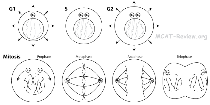
- Prophase = Prepare (condense chromatin into chromosomes, break down membrane nucléaire, assemble fuseau mitotique, centriole pairs move toward opposite poles of the cellule)
- Métaphase = Middle (Chromosomes line up in the middle)
- Anaphase = Apart (Sister chromatids pulled apart to opposite sides of cellule)
- Télophase = Prophase in reverse = de-condense chromosomes, re-form membrane nucléaire, break down fuseau mitotique.
- Mitotic structures and processes
- centrioles, asters, spindles: responsible for pulling apart the chromatides sœurs
- chromatids, centromeres, kinetochores: chromatides sœurs are duplicated copies of the chromosome. chromatids are joined at the centromere. There's a protéine at the centromere called the kinetochore, where fibres du fuseau attach to pull the chromatids apart.
- membrane nucléaire breakdown and reorganization: for most eukaryotes, the membrane nucléaire breaks down at the beginning of mitose, and reforms at the end of mitose around each of the two newly formed nuclei.
- mechanisms of chromosome movement: chromatids move apart during anaphase by the fibres du fuseau. Microtubules cause the chromosome movement.
- Phases of cycle cellulaire: G0, G1, S, G2, M
- G0 = no more réplication de l'ADN or division cellulaire. Examples include nerfs and muscles.
- G1 = growth = make organites, increase in cellule size.
- S = réplication de l'ADN. Centrioles also replicated.
- G2 = growth = make organites, increase in cellule size.
- M = mitose.
- Growth arrest: the cycle cellulaire can be arrested for many reasons:
- Too much genomic mutation/damage causes a cellule to arrest in M phase.
- Contact inhibition: normal epithelial cellules stop growing when it gets crowded such that it's touching adjacent cellules.
- Lack of food can also cause growth arrest.
- Apoptose (Programmed Cellule Death)
- Apoptose = death that is clean and healthy.
- Apoptose = activation of caspases that digest the cellule from within.
- No spilling of cellule contents.
- Afterwards, the apoptosed cellule releases chemicals that attract macrophages, and gets engulfed.
- Apoptose can be brought upon by development (eg tadpole losing tail) or by réponse immunitaire (infected/cancerous cellules killed by cellules T cytotoxiques/cellules tueuses naturelles).
      </div>

      <div class="fiche-section">
        <div class="fiche-section-title">Biosignalisation</div>
<ul class="fiche-list"> </div>
<div class="source-note">Contenu adapté de <a href="https://mcat-review.org/generalized-eukaryotic-cell.php" target="_blank" rel="noopener">mcat-review.org</a> — Traduction et mise en forme par Logopoïos</div>
</div>
</div>
<div class="chapter" id="ch-7">
<div class="chapter-header" onclick="toggleChapter(this)">
<div>
<div class="chapter-num">Chapitre 07</div>
<div class="chapter-title">Génétique</div>
</div>
<div class="chapter-toggle">▼</div>
</div>
<div class="chapter-body">
<div class="fiche-section">
<div class="fiche-section-title">Concepts mendéliens</div>
<ul class="fiche-list">- Phenotype: what is observed. For example, height, color, whether the organism exhibits a trait.
- Genotype: the genetic make up. For example, homozygous dominant (TT), heterozygous (Tt), homozygous recessive (tt).
- Gène: a gène is a stretch of DNA that codes for a trait. In molecular biology, the gène codes for a protéine, which acts to bring about a trait.
- Locus: location (of a gène) on a chromosome.
- Allèle: single and multiple
- An allèle is a variant of a gène. A gène may have a number of allèles. All allèles of the same gène exist at the same locus.
- A cellule holds 2 allèles of each gène. One allèle from mom, one allèle from dad.
- When a gène has only 2 allèles, then that's the simple case we're used to seeing. For example, the trait for height in peas is governed by T and t. TT and Tt gives tall plants, and tt gives short ones.
- When a gène has more than 2 allèles, then that's called multiple allèles. For example, groupe sanguin is governed 3 allèles: IA IB and i. Because a cellule can only hold 2 of these allèles, the different combinations an individual can have are:
GenotypeBlood type (phenotype)
IAIA or IAi
A
IBIB or IBi
B
IAIB
AB
ii
O
- Homo- and heterozygosity
- Homozygous: when the two allèles that an individual carries are the same. For example, AA or aa.
- Heterozygous: when the two allèles that an individual carry are different. For example, Aa.
- Wild type: the "normal" allèle or phenotype for an organism. The type sauvage is usually the most prevalent, although it doesn't necessarily have to be true.
- Recessiveness: the "weak" allèle. The allèle récessif is only expressed if both copies are present. Only a single copy is needed for the allèle dominant. The allèle récessif is usually denoted as the lower case letter, the allèle dominant is usually denoted as the upper case letter. For example, blond hair is recessive. Both allèles for blond hair need to be present, otherwise the hair is dark.
- Complete dominance
GenotypePhenotype
AADominant
AaDominant
aaRecessive
- Co-dominance
GenotypePhenotype
AAA
ABBoth A and B
BBB
An example of codominance is the A and B groupe sanguin allèles. Type A cellules have A antigènes. Type cellules B have B antigènes. Type AB makes both antigènes.
- Indominance complète, leakage, pénétrance, expressivité
- Indominance complète:
GenotypePhenotype
AAA
ABIn between A and B
BBB
An example of dominance incomplète is the color of chickens. A cross between black chickens and white chickens give rise to bluish grey chickens.
- leakage: gène flow from one species to another.
- Pénétrance is the frequency that a genotype will show up in the phenotype. 100% pénétrance means that if you have the gènes for being smart, then you'll definitely be smart! Less than 100% pénétrance means that you may have the gènes for being smart, but you may not actually be smart.
- Expressivité is to what degree a penetrant gène is expressed. Constant expressivité means that if your gènes for being smart manages to penetrate (show up as a trait), then you're IQ is 120. Variable expressivité means that your IQ doesn't have to be 120, it could be somewhat lower or somewhat higher.
- Hybridation: viability
- Hybrid vigor = increased viability for offspring of parents who are genetically more different
- Mechanism = less chance to receive 2 copies of the same detrimental recessive gène
- Gène pool: all of the allèles in a population.
      <div class="fiche-section">
        <div class="fiche-section-title">Méiose et variabilité génétique</div>
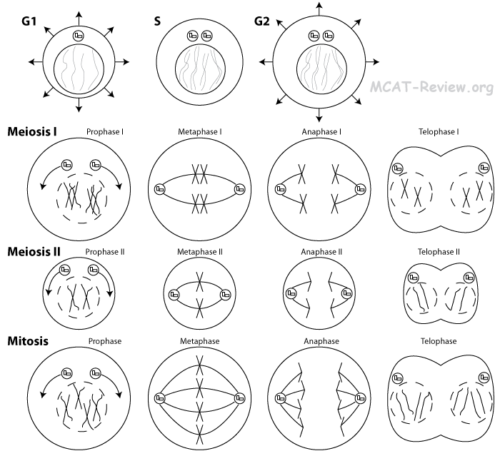
<ul class="fiche-list">- Important differences between méiose and mitose
mitose
méiose
no tetrad
tetrad formation (pairing of chromosomes homologues) and enjambement
daughter cellules identical to parent cellule
daughter cellules different from parent cellule
diploid (2n) daughter cellules
haploid (n) daughter cellules
1 division involved
2 divisions involved
2 daughter cellules
4 spermatozoïde cellules or 1 ovule (with polar bodies)
- Segregation of gènes
- Independent assortment
- Independent assortment generates variation génétique.
- A cellule has 2 copies of each somatic chromosome- one from mom, one from dad (chromosomes homologues). Independent assortment shuffles these chromosomes, and then places only one copy of each into the gamete. This way, the gamete may have chromosome 1 from mom, chromosome 2 from dad, chromosome 3 from dad, ... etc.
- The mechanism of assortiment indépendant is the following: During métaphase I of méiose, chromosome homologue pair up along the métaphase line in random orientation - sometimes the mom's chromosome is on the left, sometimes it's on the right. During anaphase I of méiose, the chromosomes homologues are pulled apart. Those on the left will be put into one daughter cellule, those on the right will be put into another.
- Linkage
- Because of assortiment indépendant, gènes on different chromosomes are randomized. However, gènes on the same chromosome can not be randomized by this mechanism.
- Gènes on the same chromosome are linked to some extent.
- Crossing over is a mechanism that reduces linkage. However, enjambement is only efficient when the gènes are physically apart from each other on the chromosome.
- When the gènes are further apart on the chromosome, enjambement makes them less linked.
- The physically closer the gènes are on the chromosome, the more linked they are.
- Recombination: also called recombinaison génétique, is the process that introduces diversité génétique into the gametes during méiose. There are 2 processes that makeup recombination: assortiment indépendant and enjambement.
- Crossing over occurs during prophase I (the actual site of enjambement is the chiasma. The chiasma is made possible because of pairing of chromosomes homologues called the tetrad, which is formed by a process called synapsis).
- Single crossovers: results in recombinaison génétique. The chromatids involved in this single crossover exchange allèles at a given locus. Results in 2/4 recombinants.
- Double crossovers:
- Scenario 1: results in no recombinaison génétique. The chromatids involved in this double crossover exchange allèles at first, but then it exchanges them back, resulting in no net recombination. This is called the 2-strand double crossover. Results in 0/4 recombinants.
- Scenario 2: results in recombinaison génétique. The chromatids exchange allèles during a crossover. Then, one of the crossover chromatid exchanges with a different chromatid. This is called the 3-strand double crossover. Results in 2/4 recombinants.
- Scenario 3: results in recombinaison génétique. The chromatids exchange, then 2 totally different chromatids on the same chromosome exchange. This is called the 4-strand double crossover. Results in 4/4 recombinants.
- Synaptnemal complex: the protéine complex that glues the tetrad together
- Tetrad: the paired chromosome homologue structure
- Sex-linked characteristics = gène for the characteristic is on the X chromosome.
- Very few gènes on Y chromosome
- The Y chromosome is very small and carries few gènes of importance.
- All the lié au sexe allèles are carried on the X chromosome.
- Sex determination: XX = female, XY = male
- Cytoplasmic/extranuclear inheritance
- Cytoplasmic inheritance = inheritance of things other than genomic DNA.
- All cellular organites, such as mitochondries, is inherited from the mother.
- Mutation
- General concept of mutation-error in séquence d'ADN
- Mutation = change in séquence d'ADN by means other than recombination.
- Types of mutations: random, traduction error, transcription error, substitution de base, inversion, addition, deletion, translocation, mispairing
- Random mutation = random changes in séquence d'ADN. Can be due to rayonnement, chemicals, réplication error ...etc.
- Traduction error = even if the DNA for a gène is perfect, errors during traduction can cause expression of a mutant phenotype.
- Transcription error = even if the DNA of a gène is perfect, errors during transcription can cause expression of a mutant phenotype.
- Base substitution = mutation involving a base (ATGC) changing to a different base.
- Inversion = a stretch of DNA (a segment of a chromosome) breaks off, then reattaches in the opposite orientation.
- Addition = also called insertion = an extra base is added/inserted into the séquence d'ADN.
- Deletion = a base is taken out of the séquence d'ADN.
- Addition/insertion and deletion mutations result in a mutation par décalage du cadre de lecture.
- Translocation = a stretch of DNA (a segment of a chromosome) breaks off, then reattaches somewhere else.
- Mispairing = A not pairing with T, or G not pairing with C.
- Advantageous vs. deleterious mutation
- Advantageous = results in a benefit to the fitness of the organism. For example, the mutation that causes flies to become wingless is advantageous in an environment that is very windy.
- Deleterious = results in a harmful effect to the fitness of the organism. For example, a mutation that causes an organism to be sterile.
- Inborn errors of métabolisme = maladies génétiques resulting in faulty métabolisme. For example PKU (Phenylketonuria) is an inborn error of métabolisme where people can't metabolize phenylalanine. There's no cure, but the treatment involves avoiding things containing the acide aminé phenylalanine.
- Relationship of mutagens to carcinogens
- Mutagen = something that causes mutation.
- Carcinogen = something that causes a mutation that causes cancer.
- All carcinogens are mutagens.
- Not all mutagens are carcinogens.
- Genetic drift = random changes in fréquence allélique due to chance = not due to sélection naturelle
- Synapsis or enjambement mechanism for increasing diversité génétique
- Synapsis: chromosomes homologues come together and form tetrad
- Crossing-over: exchange of matériel génétique between chromosomes homologues after the tetrad forms
- Increases diversité génétique by introducing new allèle combinations in the chromosome you're about to give to your offspring
      </div>

      <div class="fiche-section">
        <div class="fiche-section-title">Méthodes analytiques</div>
<ul class="fiche-list">- p+q = 1
- (p+q)2 = 1 → p2 + 2pq + q2 = 1
- Five Assumptions of Hardy-Weinberg
- Infinitely large population (no dérive génétique)
- No mutation
- No migration
- Random mating (no sélection sexuelle)
- No sélection naturelle
- Test cross: rétrocroisement, concepts of parental, F1 and F2 generations
- Test cross: so you have something with dominant phenotype. It could either be Aa or AA. To find out, you cross it with the homozygous recessive aa. If Aa, half the offspring will express the recessive phenotype. If AA, no offspring will express the recessive phenotype.
- Back cross = mating between the offspring and the parent = preserve parental genotype.
- Parental generation = P = generation of the parent. On a pedigree, the is the row that represents the parents
- F1 generation = Felial 1 = children. On a pedigree, this is the row below the parents, and represents the children of the parents.
- F2 generation = Felial 2 = grandchildren. On a pedigree, this is the row below the F1, and represents the children of the F1 and grandchildren of the parents.
- Gène mapping: crossover frequencies
- Gène mapping = physical location gènes on the chromosome (eg. toward the end, closer to the middle, etc)
- Further apart = higher crossover frequency
- Biometry: statistical methods
- Biometry = using statistics to analyze biological data
- null hypothesis = first assuming that you are wrong - there is no relationship in your data
- p value = calculated chance that the null hypothesis is right, that you are wrong
- p value
      </div>

      <div class="source-note">Contenu adapté de <a href="https://mcat-review.org/genetics.php" target="_blank" rel="noopener">mcat-review.org</a> — Traduction et mise en forme par Logopoïos</div>
    </div>
  </div>

  <div class="chapter" id="ch-8">
    <div class="chapter-header" onclick="toggleChapter(this)">
      <div>
        <div class="chapter-num">Chapitre 08</div>
        <div class="chapter-title">Microbiologie</div>
      </div>
      <div class="chapter-toggle">▼</div>
    </div>
    <div class="chapter-body">

      <div class="fiche-section">
        <div class="fiche-section-title">Théorie cellulaire</div>
<ul class="fiche-list">- Living things are made of cellules
- The cellule is the most basic unit
- Cellules divide and self-replicate
- History: cellules were first discovered by Robert Hooke using a microscope to examine cork (wood)
- Impact: promoted the study of cellules and the field of microbiology and cellule biology
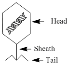
      <div class="fiche-section">
        <div class="fiche-section-title">Classification et structure des cellules procaryotes</div>
<ul class="fiche-list">- Lack of typical eukaryotic organites: Bacteria don't have Golgi, ER, mitochondries, chloroplasts.
- Prokaryotic domains
- Eubacteria are the bacteria we encounter every day
- Archaea are the prokaryotes that inhabit extreme environments (high salt, temperature, or chemicals).
- Major classifications of bacteria by shape
- Bacilli (rod-shaped): Streptococcus causing pneumonia, strep throat
- Spirilli (spiral shaped): Helicobacter pylori causing gastric ulcers
- Cocci (spherical): Staphylococcus causing skin infections/cellulitis/MRSA
- Presence of paroi cellulaire in bacteria: bacterial paroi cellulaire is made of peptidoglycan, a polysaccharide-protéine molecule. In contrast, plant paroi cellulaire is made of cellulose and fungi paroi cellulaire is made of chitin.
- Flagellar propulsion, mechanism
- Bacterial flagella is made of flagellin. In contrast, eukaryotic flagella is made of microtubules.
- The mechanism of the bacterial flagella is rotation. A rotor at the base of the flagella drives the rotation, powered by a proton or sodium gradient. (Compare this to eukaryotic flagella, which is powered directly by ATP)
      </div>

      <div class="fiche-section">
        <div class="fiche-section-title">Cellule procaryote : croissance et physiologie</div>
<ul class="fiche-list">- DNA replicates
- Replicated DNAs separate by attaching to the membrane cellulaire as the cellule elongates (in contrast to mitose, no fibres du fuseau needed).
- Cytocinèse divides the parent cellule into two daughter cellules.
- High degree of genetic adaptability, acquisition of antibiotic resistance
- Mutation
- Transformation: bacteria take in plasmids and fragments d'ADN and integrates them into the genome.
- Transduction: bacteriophages undergoing lysogenic life cycle incorporate the viral DNA into the bacterial genome.
- Conjugaison: Bacteria transfer DNA between one another through the sex pilus.
- Exponential growth: Bacterial growth starts off being exponential because of the nature of fission binaire. Later, when food becomes short, and it gets crowded, growth slows and eventually plateaus.
- Existence of anaerobic and aerobic variants
- Obligate aerobe = must have oxygen for growth.
- Obligate anaerobe = dies when oxygen is present.
- Facultative anaerobe = doesn't need oxygen for growth, but grows better with oxygen.
- Symbiotic relationships
- Parasitic = bacteria benefits at the expense of the host. Disease causing bacteria are examples of parasitic relationships.
- Mutualistic = both bacteria and host benefits. For example, the E. Coli in your gut; the natural flora on your skin.
- Commensalistic = one benefits while the other has no effect.
- Chemotaxis: bacteria don't have eyes, so they sense chemicals for navigation (eg: moving toward places with higher concentrations of food, avoiding places with higher concentrations of toxins)
      </div>

      <div class="fiche-section">
        <div class="fiche-section-title">Cellule procaryote : génétique</div>
<ul class="fiche-list">- Plasmids are double brin DNA.
- A plasmid can exist and replicate independently of the genomic DNA, or be integrated into it.
- Plasmids are inherited.
- Plasmids are not essential for growth and reproduction in the wild.
- Conjugaison transfers matériel génétique between bacteria via a pillus.
- A bacteria able to make the pillus (F+) has a plasmid that contains the pillus gènes.
- F+ bacteria can transfer the plasmid to an F- bacteria.
- Conjugaison can also transfer some genomic DNA (because F+ plasmid can integrate into the chromosome).
- Transformation: incorporation into bacterial genome of fragments d'ADN from external medium
- When a bacteria dies, it lyses and spills many fragments d'ADN into the environment.
- Another bacteria encounters these fragments d'ADN, takes them in, and integrates them into its own genome.
- If the fragments d'ADN contained an antibiotic resistant gène, then the transformation just made the bacteria antibiotic resistant.
- Régulation of expression génique, coupling of transcription and traduction
- Régulation at the transcription level: some gènes are actively transcribed, while others are not. Activateurs and inhibiteurs modulate the transcription of a gène.
- Régulation at the traduction level: Some mRNA gets translated more. In prokaryotes, mRNAs with better Shine-Dalgarno sequence are translated more. In eukaryotes, traduction régulation can involve adding more polyAs to mRNA (longer mRNA life time), modulating the traduction machinery (phosphorylation of initiation factors), or storing mRNAs to be translated at a later time (mRNA masking).
- Prokaryotes regulate expression génique predominantly at the transcription level (eg. Operons, in which inducers increase transcription, and inhibiteurs decrease transcription). Eukaryotes have more régulation at other levels, and can also undergo épissage de l'ARN, which can splice RNA in different ways to make different mRNAs.
- Transcription-traduction coupling: in prokaryotes, traduction occurs as the mRNA is being transcribed (no maturation de l'ARN in prokaryotes).
- In a coupled transcription-traduction system, régulation by attenuation can occur for the Trp gène:
- When cellule is full of Trp, traduction occurs fast because of abundant Trp acide aminé. This fast ribosome movement across the transcribing mRNA causes the Trp mRNA transcription to terminate. Because Trp is not needed.
- When cellule is starved of Trp, traduction occurs slower because Trp acide aminé is lacking. This slower ribosome movement across the transcribing mRNA causes the Trp mRNA to be made to its completion.
      </div>

      <div class="fiche-section">
        <div class="fiche-section-title">Structure des virus</div>
<ul class="fiche-list">- Nucleic acid can be DNA or RNA, simple brin or double brin.
- Protéine coat covers the acide nucléique.
- Some viruses have an envelope derived from the host's membrane cellulaire, while others lack it (nonenveloped).
- Enveloped viruses bud off the host's membrane.
- Nonenveloped viruses cause the host to burst to release viral particles.
- Smaller than bacteria.
- Lack organites, noyau: Viruses don't have any organites or a noyau. The matériel génétique is simply packed inside a capside protéique.
- Structural aspects of typical bacteriophage
- Head stores matériel génétique.
- Sheath provides a passage way for matériel génétique to be injected into the host bacteria.
- Tail fibers attach to the host bacteria.
- Genomic content RNA or DNA: Viruses can contain either RNA or DNA as their genomic content. Out of the RNA viruses, those that convert their genome into DNA inside their host are called retroviruses.
- Size relative to bacteria and eukaryotic cellules: Viruses are roughly 100 times smaller than bacteria, and 1000 times smaller than eukaryotic cellules.
      </div>

      <div class="fiche-section">
        <div class="fiche-section-title">Cycle de vie viral</div>
<ul class="fiche-list">- Generalized phage and animal virus life cycles:
- attachment to host, penetration of membrane cellulaire or paroi cellulaire, and entry of viral matériel génétique
- use of host synthetic mechanism to replicate viral components: Host's ribosomes synthesize the necessary enzymes. Host's ATP provides necessary energy. The host also provides the raw materials such as nucleotides and acides aminés.
- self-assembly and release of new viral particles: The coat protéines and viral matériel génétique will assemble into viral particles all by themselves.
- Retrovirus life cycle: integration into host DNA
- First, retrovirus enters the host.
- The viral reverse transcriptase then converts the viral RNA genome into double brin DNA.
- A virally encoded enzyme called integrase adds in the viral DNA into the host's genome at a random place.
- When the host replicates, the viral DNA gets replicated also.
- Transduction: transfer of matériel génétique by viruses
- Virus infects cellule: host DNA degraded into fragments, viral DNA takes over control.
- Host fragment d'ADN gets packed into virus progeny by accident.
- Virus progeny infects another cellule, injects previous host's fragment d'ADN.
- Fragment enters cellule, find its homologous counterpart, and crossover.
      </div>

      <div class="fiche-section">
        <div class="fiche-section-title">Champignons</div>
<ul class="fiche-list">- Made of hyphae filaments.
- Parasitic hyphae = haustoria
- A mass of hyphae is called mycelium.
- Have paroi cellulaire made of chitin.
- All fungi are heterotrophs - they are either parasites or saprobes.
- Lichens = fungi + algae. Algae provides food, fungi provides water and protection.
- Mycorrhizae = fungi + plant roots. Plant provides food, fungi provides more absorption surface.
- Yeast, molds, mushrooms are all fungi.
- General aspects of life cycle
- Can be sexual or asexual.
- Reproduces via spores or mycelial fragmentation.
- Most fungi have both a haploid and a diploid stage of life cycle.
</div>
<div class="source-note">Contenu adapté de <a href="https://mcat-review.org/microbiology.php" target="_blank" rel="noopener">mcat-review.org</a> — Traduction et mise en forme par Logopoïos</div>
</div>
</div>
<div class="chapter" id="ch-9">
<div class="chapter-header" onclick="toggleChapter(this)">
<div>
<div class="chapter-num">Chapitre 09</div>
<div class="chapter-title">Biologie moléculaire : Structure et fonction de l'ADN</div>
</div>
<div class="chapter-toggle">▼</div>
</div>
<div class="chapter-body">
<div class="fiche-section">
<div class="fiche-section-title">Structure et fonction des acides nucléiques</div>
<ul class="fiche-list">- DNA = deoxy-ribo-acide nucléique
- RNA = ribo-acide nucléique
- deoxy = 2'-H (vs 2'-OH in RNA)
- ribo = ribose sugar = pentose sugar
- acid = phosphate group gives it acidity
- Nucleotides and nucleosides
- Nucleotide = base (Adenine, Guanine, Thymine, Cytosine) + sugar + phosphate.
- Nucleoside = base + sugar = Adenosine, Guanosine, Thymidine, Cytidine.
- Sugar phosphate backbone = phosphodiester bonds linking the 5' phosphate to the 3' hydroxyl group of the pentose sugar
- Base can either be purines A and G (the big ones with 2 rings) or pyrimidines T and C (the small ones with 1 ring).
- Thymine is replaced with Uracil in RNA
- The phosphate group gives DNA its acidity.
- Deoxyriboacide nucléique (DNA): double helix, Watson-Crick model of DNA structure
- The "double" in the double helix means that DNA is found in a double brin form - 2 simple brin chains of DNA stuck to each other via liaison hydrogène of the paires de bases.
- The 2 single-strands are anti-parallel to each other. Going from 5' to 3' of one strand means going from 3' to 5' of the other strand.
- The "helix" in the double helix means that the entire thing is wound up in a spiral.
- Base pairing specificity: A with T, G with C
- A forms 2 liaisons hydrogène with T.
- G forms 3 liaisons hydrogène with C.
- GC bonds are stronger. DNA with high GC content will be harder to break apart.
- Complementary strands of DNA liaison hydrogène with each other.
- 5'-ATGC-3' will be complementary to 5'-GCAT-3' or 3'-TACG-5', but NOT 5'-TACG-3'. make sure you get the 5's and 3's right.
- Fonction in transmission of genetic information
- Because of the complementary nature of paire de basesing, DNA can transmit genetic information through réplication.
- DNA dénaturation, reannealing, hybridation
- dénaturation = separation of 2 brins complémentaires = caused by high temperature
- reannealing = coming together of the 2 brins complémentaires after they've been separated
- hybridation = annealing = coming together of brins complémentaires
- annealing is generally used in PCR reactions, where primer anneals to the brin matrice
- hybridation is generally used in Southern blotting (probe anneals to target) and plasmid ligation (sticky ends anneal)
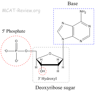
<div class="fiche-section">
<div class="fiche-section-title">Réplication de l'ADN</div>
<ul class="fiche-list">- First, the double brin DNA must separate, or unwind. To do this:
- DNA gyrase (class II topoisomerase) is responsible for uncoiling the DNA ahead of the réplication fork.
- Helicase is responsible for unwinding the DNA at the réplication fork.
- Single-strand binding protéine (SSB) is responsible for keeping the DNA unwound after the helicase. SSBs stabilize simple brin DNA by binding to it.
- Next, you start making DNA that is complementary to the newly unwound/separated DNA. Note, all biological synthèse d'ADN occurs from the 5' to the 3' end.
- Primase gets this started by laying down a short RNA primer on the unwound DNA. The primer is made of RNA, but is complementary to the séquence d'ADN. Later, this RNA is replaced with DNA.
- ADN polymérase then takes over and makes DNA that is complementary to the unwound DNA.
- synthèse d'ADN occurs on both strands of the unwound DNA. The synthesis that proceeds in the direction of the réplication fork is the brin directeur. The synthesis that proceeds in the opposite direction to the réplication fork is the brin retardé. The brin retardé contains Okazaki fragments.
- Finally, RNA primers are replaced with DNA by a special ADN polymérase. The Okazaki fragments in the brin retardés are then stitched together by DNA ligase.
- synthèse d'ADN is bidirectional: 2 réplication forks form and proceeds in opposite directions (like an expanding bubble).
- Biological synthèse d'ADN always proceeds from the 5' end to the 3' end.
- ADN polymérase has proof-reading activity, which means it corrects any mistakes (mutations) it makes.
- Réplication occurs once every cellule generation, during the S phase. (Cellule division may occur twice in méiose, but réplication still occurs once only)
- Semi-conservative nature of réplication
- Newly synthesized DNA contains one old strand and one new strand.
- Meselson and Stahl proved this by experiment: Basically, they used heavy (15N) DNA as the old (pre-réplication) DNA, and used light (14N) nucleotides for the synthesis of new DNA. They can tell the difference between heavy and light DNA by centrifugation. What they found was that when heavy DNA undergoes one round of réplication in light nucleotides, the DNA made is of intermediate weight. After the second round of réplication, the DNA is split between intermediate and light weight.
- If réplication de l'ADN were completely conservative, only heavy and light DNA would be seen, and nothing in between. This was not the case.
- If réplication de l'ADN were dispersive, everything would be of intermediate weight. Again, this was not the case because after the second round of réplication, light DNA was seen.
<div class="fiche-section">
<div class="fiche-section-title">Réparation de l'ADN</div>
<ul class="fiche-list">- ADN polymérase has proof-reading activity (also called 3' → 5' exonuclease activity). If a wrong nucleotide gets incorporated, the polymerase will "back-up" and replace it with the correct one.
- The special polymerase that replaces the RNA primers with DNA also have 5' → 3' activity. This allows the polymerase to clear away short stretches of incorrect nucleotides (RNA or incorrect DNA) and replace it with the right ones (DNA). This process is also called repair.
- Repair of mutations
- Mismatch repair: enzymes recognize incorrectly paired paires de bases and cuts out the stretch of DNA containing the mismatch. Then polymerase re-adds the correct nucleotides in.
- During mismatch repair, the repair enzyme must decide what strand of DNA to cut since DNA contains 2 strands. To do this, the enzyme cuts the DNA strand that do not have methylations. The original (old) DNA has methylations, but the newly synthesized DNA do not have them until shortly after réplication. Thus, there is a window of time when mismatch repair enzymes can know what strand to cut if mismatch is encountered.
- Base-excision repair: a damaged base gets cut out. Then the base's sugar phosphate backbone gets cut out. And then, several more nucleotides next to the base get cut out. Finally, polymerase remakes the cut out nucleotides.
- Nucleotide-excision repair: damaged nucleotide(s) gets cut out and then polymerase replaces it. This is like mismatch-repair, but it's not for mismatch. It's for damages like thymine dimers, and other damages that changes normal nucleotides into abnormal nucleotides.
- Nick traduction: this is basically 5' → 3' exonuclease activity coupled to polymerase activity. The polymerase here chugs along, chews off the bad nucleotides and then replaces them with new nucleotides. This is what happens when RNA primers are replaced with DNA.
- SOS response in E. Coli: during réplication, when there's just too much dommages à l'ADN for normal repair to handle, the SOS repair system comes along. Instead of correcting any dommages à l'ADNs during réplication, the polymerase replicates over the damaged DNA as if it were normal. By using the damaged DNA as a template error rates are high, but it's still better than not replicating at all.
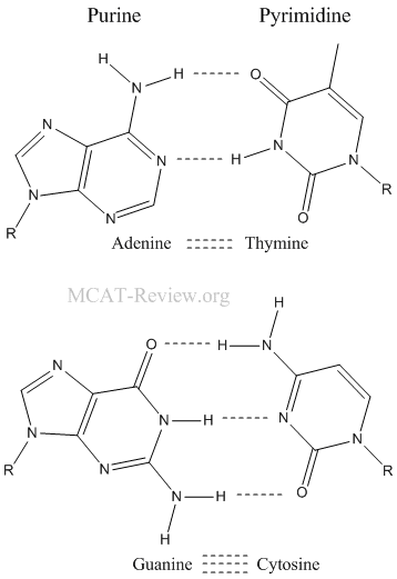
      <div class="fiche-section">
        <div class="fiche-section-title">ADN recombinant</div>
<ul class="fiche-list">- The plasmid must have a restriction site because you need to open it up for the insertion of your gène.
- The plasmid must have an origin of réplication because you want to clone your gène, which is inside your plasmid.
- The plasmid must have an antibiotic resistant gène because this lets you kill competing, useless bacteria that doesn't have your plasmid. When you add an antibiotic, only the bacteria with the antibiotic resistant plasmid will live.
- Plasmids replicate independently of the genomic DNA of the bacteria.
- Restriction enzymes
- Restriction enzymes (also called restriction endonucleases) cut double brin DNA at palindrome sequences. The resulting fragments are called restriction fragments.
- If you read from 5' → 3' of one strand, then read from 5' → 3' of the other strand, and they are the same, then the section of the double brin DNA that you just read is a palindrome sequence.
- Some restriction enzymes cut to make sticky ends, which can hybridize.
- Some restriction enzymes cut to make blunt ends, which cannot hybridize.
- DNA libraries = the result of cloning an organism's entire DNA (genomic or cDNA)
- Genomic library = entire genomic DNA -> fragmented by restriction enzyme -> cloned
- cDNA library = entire mRNA population -> reverse transcription into cDNA -> cloned
- Generation of cDNA: mRNA + reverse transcriptase = cDNA
- Hybridation
- Hybridation, also called annealing, is where DNA strands paire de bases with each other.
- In Southern blotting, DNA probes are used to hybridize onto fragments d'ADN containing a target sequence.
- In clonage de gènes, hybridation refers to the process where sticky ends from a restriction fragment of a gène paires de bases with the same sticky ends on a plasmid.
- Expressing cloned gènes
- Expression = Cloned gène -> mRNA -> protéine
- Expression needs a vector with the transcription and traduction machinery
- The cloned gène needs to be placed downstream of a strong promoter so that a lot of mRNA will be transcribed
- PCR
- Dénaturation: heat (90 °C) to separate double brin matrice d'ADN.
- Annealing: cool reaction in order for primers to anneal to the now simple brin matrice d'ADN.
- Excess amount of primers, so they out complete re-annealing of the brin matrices.
- Elongation: use heat stable polymerase to extend the primers.
- Repeat steps 1 to 3 for n cycles. The resulting amplification of the original matrice d'ADN after n cycles is 2n.
- Gel electrophoresis and Southern blotting
- Gel electrophoresis = separates fragments d'ADN on a sheet of gel
- Southern blotting = probes make your DNA light up on that sheet of gel
- Probe for southern blotting = DNA that is complementary to the target DNA
- Other blots
- Northern blot: RNA target, DNA/RNA probe
- Western blot: protéine target, anticorps probe
- DNA sequencing
- Sanger sequencing
- basis: differential chain termination (using dideoxy-NTPs), then detected by capillaire electrophoresis
- pros: cheap
- cons: can only sequence a short distance, can only detect mutations if it's at high levels
- Nextgen sequencing = massive parallel sequencing
- basis: addition of different nucleotide causes different color fluoresence, detected on a 2D chip containing many simultaneous reactions
- pros: can sequence an entire genome, can detect mutations at very low levels
- cons: expensive
- Analyzing expression génique
- Gène expression correlates with mRNA levels, so measure RNA
- What gènes are expressed and at what levels (global picture)? Use expression génique profiling (microarray). Eg: what gènes are overexpressed in certain cancer
- How much is this gène expressed? Use reverse transcription then real time quantitative PCR. Eg: residual BCR-ABL in CML
- Stem cellules
- Stem cellules = cellules that have the potential to differentiate into many different types cellulaires
- Example: hematopoietic cellules souches -> neutrophils, lymphocytes, globules rouges, platelets
- Uses: hematopoietic cellule souche (moelle osseuse) transplant as a cure for leukemia
- Practical applications of DNA technology
- Medical applications: PCR coupled with DNA sequencing = detects mutations / variants = used for diagnosis, monitoring disease level, or predict whether a patient will respond to a certain treatment
- Human thérapie génique = using viruses to infect and introduce functional gènes into human cellules
- Pharmaceuticals: uses cloning, expression, and protéine purification to isolate protéines/enzymes to treat patients who are deficient in those
- Forensic evidence: DNA fingerprinting = paternity test = identifies a criminal/person
- Environmental cleanup: they are making génétiquement modifié bacteria that can breakdown oil spills or plastic polymers
- Agriculture: génétiquement modifié foods = introducing gènes in crops to make them grow better and be resistant to pests
- Safety and ethics of DNA technology
- Side effect of human thérapie génique: the viruses make mistakes when introducing the gènes, resulting in mutations that cause cancer
- We are not absolutely certain about the long term health effects of génétiquement modifié foods
- Sequencing someone's entire genome exposes all their underlying maladies génétiques - potential basis for discrimination from insurance companies
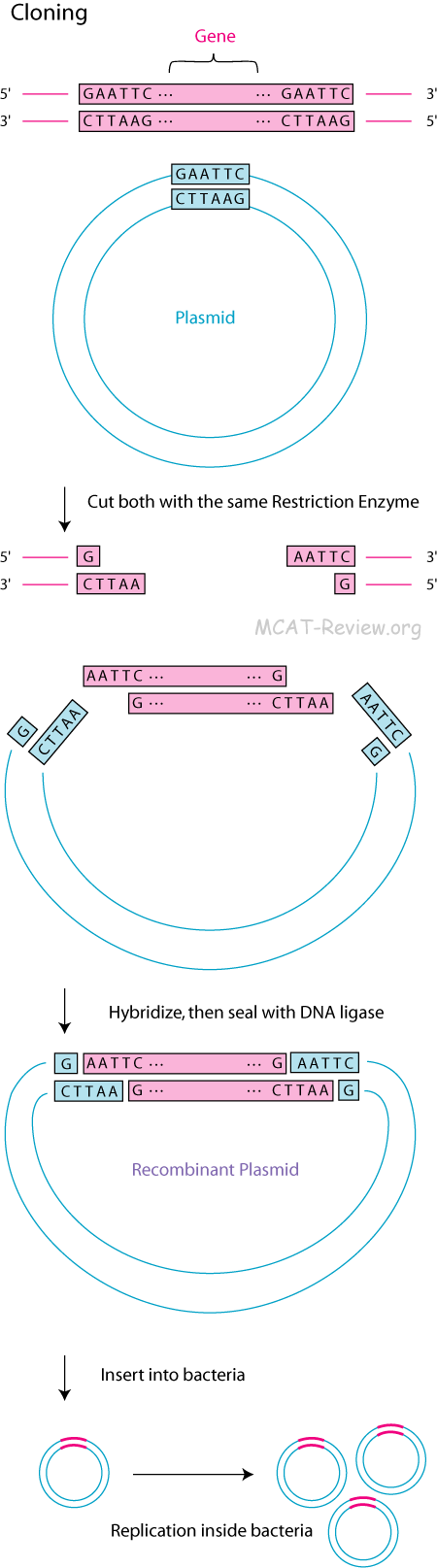
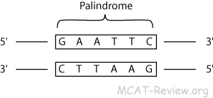
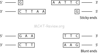
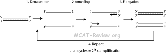
      <div class="source-note">Contenu adapté de <a href="https://mcat-review.org/molecular-biology-dna.php" target="_blank" rel="noopener">mcat-review.org</a> — Traduction et mise en forme par Logopoïos</div>
    </div>
  </div>

  <div class="chapter" id="ch-10">
    <div class="chapter-header" onclick="toggleChapter(this)">
      <div>
        <div class="chapter-num">Chapitre 10</div>
        <div class="chapter-title">Biologie moléculaire : Eucaryotes</div>
      </div>
      <div class="chapter-toggle">▼</div>
    </div>
    <div class="chapter-body">

      <div class="fiche-section">
        <div class="fiche-section-title">Organisation des chromosomes eucaryotes</div>
<ul class="fiche-list">- Histones: responsible for the compact packing and winding of chromosomal DNA. DNA winds itself around histone octamers.
- nonhistone chromosomal protéines: all the other protéines are lumped together in this group. Responsible for various roles, such regulatory and enzymatic.
- Single copy vs repetitive DNA
- Tandem repeats (repeats right next to each other, eg: CAGCAGCAG)
- Trinucleotide repeats: too much can cause diseases. Eg: Huntington disease and fragile X syndrome
- Satellites, minisatellites and microsatellites: unique pattern of repeats identifies an individual (paternity testing, forensics) or a particular transplant donor (moelle osseuse engraftment studies)
- Interspersed repeats: mobile elements/transposons have repetitive DNA at both ends of the sequence
- Supercoiling: a coil on top of a coil, helps to compact very large amounts of genomic DNA to fit into a compact chromosome
- Telomeres, centromeres
- Telomere: the 2 ends of the chromosome.
- Centromere: a region on the chromosome, can be at the center or close to one of the ends. After réplication, chromatides sœurs are attached at the centromere. During mitose, fibres du fuseau are attached at the centromere and pulls the chromatides sœurs apart.
- A common question is what is the difference between chromatin and chromosome. The answer is chromatin is the "stuff" that chromosomes are made of. If the chromosome is a cotton shirt, then chromatin is cotton.
</div>
<div class="fiche-section">
<div class="fiche-section-title">Contrôle de l'expression génique chez les procaryotes</div>
<ul class="fiche-list"> </div>
<div class="fiche-section">
<div class="fiche-section-title">Contrôle de l'expression génique chez les eucaryotes</div>
<ul class="fiche-list">- Transcription factors (protéine) bind to enhancers or silencers (DNA) to affect transcription. Enhancers increase transcription when bound, while silencers decrease it. The main difference in eukaryotes that sets them apart from prokaryotes is that enhancers/silencers can be very far away from the actual promoter, and can be upstream or downstream. The DNA must must loop back on itself so that the facteur de transcription bound to enhancer/silencer can actually make contact with the promoter. Intermediate protéines are involved in the process.
- Eukaryotes lack the bacterial transcription régulation mechanisms such as the operon and attenuation.
- DNA binding protéines, facteurs de transcription
- DNA-binding protéines bind to DNA.
- facteurs de transcription bind to DNA, so they have a DNA-binding domain.
- DNA-binding domains interact with the grooves in the double helix (major grooves and minor grooves).
- Advanced: common DNA-binding domains include helix-turn-helix (HTH), zinc finger, basic-region leucine zipper (bZIP).
- Gène amplification and duplication
- Gène duplication = 2x amplification
- Mechanism: either a portion of DNA gets duplicated within a chromosome or the entire chromosome is duplicated
- Certain gènes are amplified in cancer such as MYC and RAS
- Certain amplification de gèness can be used as a target for treatment: Her2 amplification is treated with Herceptin in breast cancer
- Certain amplification de gèness affect the prognosis of a disease: intrachromosomal amplification of chromosome 21 = worse prognosis in acute lymphoblastic leukemia
- Other amplification de gèness have no effect at all other than to increase the size of the genome (eg. Selfish/parasitic DNA that serves no function but is amplified)
- Post-transcriptional control, basic concept of splicing (introns, exons)
- tRNAs and rRNAs modifications: some normal nucleotides are modified to control the structure of these RNAs.
- mRNAs modifications
- épissage de l'ARN: sequences called introns are cut out, sequences called exons are kept and spliced (joined) together.
- Alternate splicing: different ways of cutting up and RNA and rejoining the exons pieces make different final RNA produits.
- 5' capping and 3' poly-A tail: these help to protect the RNA from dégradation so they can last longer.
- After the correct modifications, RNA is transported out of the noyau where they can function in traduction.
- After some time, RNA is degraded. The rate and timing of RNA dégradation can be controlled by the cellule.
- Cancer as a failure of normal cellular controls, oncogenes
- Failure of normal cellular controls:
- Cancer cellules continue to grow and divide in situations normal cellules would not.
- Cancer cellules fail to respond to cellular controls and signals that would halt this growth in normal cellules.
- Cancer cellules avoid apoptose (self-destruction) that normal cellules undergo when extensive dommages à l'ADN is present.
- Cancer cellules stimulate angiogenesis (cause new vaisseaux sanguins to grow to nourish the cancer cellule).
- Cancer cellules are immortal while normal cellules die after a number of divisions.
- Cancer cellules can metastasize - break off and then grow in another location.
- Oncogenes: gènes that cause cancer when activated. The produit of many oncogenes are involved in speeding up division cellulaire. Before an oncogene is activated, it is a harmless proto-oncogene. Something occurs that changes the proto-oncogene to an oncogene. The classic example of oncogene is the src.
- Tumor suppressors: if the oncogene is the "bad" gène, tumor suppressors are the "good" gènes. The produit of many tumor suppressors are involved in slowing down or controlling division cellulaire. If something happens that cause the tumor suppressor to no longer function, then the cellule becomes cancerous. The classic example of tumor suppressor is the p53.
- Régulation of chromatin structure
- Chromatin = DNA wrapped around histones
- Chromatin can be regulated by modifying DNA (méthylation) or histone (méthylation, acétylation)
- Euchromatin: less compact chromatin, more exposed DNA, actively transcribed
- Heterochromatin: more compact chromatin, less exposed DNA, not actively transcribed
- DNA méthylation = CpG méthylation (natural) or alkylating agent (chemotherapy) induced méthylation
- CpG islands = GC-rich séquence d'ADN near promoters that is the target for méthylation
- The méthylation occurs on the cytosine nucleotide to make 5-methylcytosine
- The enzyme is DNA methyltransferase (DNMT)
- Methyltransferases can work in both ways (add or remove methyl groups), depending on the enzyme
- Methylating CpG islands silences a gène
- Clinical utility: MGMT promotor méthylation status
- Glioblastoma (cerveau cancer) is treated by an alkylating agent (chemotherapy) +/- rayonnement
- MGMT = methyltransferase enzyme that removes alkyl/methyl groups from DNA = undo's the chemotherapy
- Bad = active MGMT = unmethylated MGMT promotor = undo's chemotherapy
- Good = Inactive MGMT = methylated MGMT promotor = effective chemotherapy
- Non-coding RNAs = not translated into protéine = anything other than an mRNA
- rRNA = makes up the ribosome
- tRNA = brings acides aminés to the ribosome
- snRNA = makes up the épissage de l'ARN machinery
- snoRNA = guides RNA modification
- RNA is heavily modified in RNAs where a structural purpose is needed (rRNA, tRNA, snRNA)
- 2'O méthylation (protects against hydrolysis/dégradation)
- Convert Uracil to pseudoUracil (plays a role in tRNA structure and integrity)
- miRNA = makes up the RNA silencing machinery (blocks mRNA traduction or degrades mRNA)
      </div>

      <div class="source-note">Contenu adapté de <a href="https://mcat-review.org/molecular-biology-eukaryotes.php" target="_blank" rel="noopener">mcat-review.org</a> — Traduction et mise en forme par Logopoïos</div>
    </div>
  </div>

  <div class="chapter" id="ch-11">
    <div class="chapter-header" onclick="toggleChapter(this)">
      <div>
        <div class="chapter-num">Chapitre 11</div>
        <div class="chapter-title">Biologie moléculaire : Synthèse des protéines</div>
      </div>
      <div class="chapter-toggle">▼</div>
    </div>
    <div class="chapter-body">

      <div class="fiche-section">
        <div class="fiche-section-title">Code génétique</div>
<ul class="fiche-list">- DNA: resides in the noyau. It codes information in gènes.
- Transcription: Inside the noyau, the DNA gènes get transcribed into RNA (messenger RNAs or mRNAs).
- RNA: The mRNAs get transported out of the noyau into the cytoplasme. mRNAs are working copies of the gène.
- Traduction: ribosomes read off the mRNAs to make protéines.
- Protéine: synthesized by ribosomes. They are the end produit of what's encoded in the gènes and they perform all the functions in the cellule.
- The triplet code
- mRNA (nucleotides) must code for protéine (acides aminés)
- To do this: 3 nucleotide = 1 codon = 1 acide aminé
- Codons are continuous, non-overlapping and degenerate
- Continuous because one codon follows right after another. There're no nucleotides in between
- Non-overlapping because the 3 nucleotides that consist of one codon never serve as part of another codon
- Degenerate because more than one codon codes for a given acide aminé
- Codon-anticodon relationship
- Anticodon: the 3 bases on the "tip" of the tRNA. A single tRNA contains a single anticodon at the "tip" and the corresponding acide aminé at the "tail". Anticodons are complementary to their corresponding codon.
- The codon-anticodon relationship: During traduction, codons pair with anticodons so that the correct acides aminés can be linked to a given codon.
- Degenerate code, wobble pairing
- Degenerate = more than one codon codes for a given acide aminé
- Wobble pairing = the last/3rd nucleotide of the codon doesn't have to match exactly with the anticodon, will still incoporate the same acide aminé during traduction
- Wobble pairing is the mechanism that explains the degenerate nature of the triplet code
- Missense, nonsense codons
- Missense codon: mutated codon that results in a different acide aminé
- Nonsense codon: mutated codon that results in something other than an acide aminé. For example, a codon stop
- Initiation, codon de terminaisons (function, codon sequences)
- Initiation codon (AUG): signals the start of traduction. Lies just downstream of the Shine Dalgarno sequence (Kozak sequence for eukaryotes)
- Termination codon (UAG,UGA,UAA): signals the end of traduction. Unlike other codons, tRNA are not involved. Instead a protéine called "facteur de libération" comes along and terminates traduction
- mRNA composition and structure (RNA nucleotides, 5' cap, poly-A tail)
- mRNA stands for messenger RNA. It's the produit of transcription and the template for traduction.
- The 5' cap is a modified nucleotide linked in a special way to the mRNA. This protects the 5' end from exonuclease dégradation.
- The poly-A tail protects the 3' end of the mRNA from exonuclease dégradation.
- Eukaryotic mRNA: 5' cap - nucleotides - 3' polyA.
- Prokaryotic mRNAs don't have the 5' cap or polyA tail.
- More mRNA = more level of expression = more protéine made
- Stronger promoter, less DNA/histone méthylation = more mRNA made
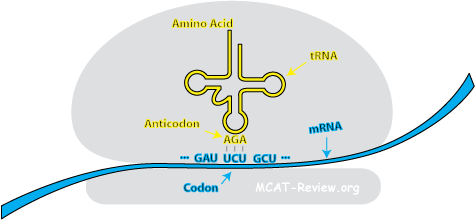
      <div class="fiche-section">
        <div class="fiche-section-title">Transcription</div>
<ul class="fiche-list">- tRNA, rRNA composition and structure (eg., RNA nucleotides)
- Both tRNA (transfer RNA) and rRNA (ribosomal RNA) are produits of transcription. However, they do not serve as the template of traduction. tRNA is responsible for bringing in the correct acide aminé during traduction. rRNA makes up the ribosome, which is the enzyme responsible for traduction.
- tRNA is made of nucleotides, many of which is modified for structural and functional reasons. At the 3' end of the tRNA, the acide aminé is attached to the 3'OH via an ester linkage.
- tRNA structure: clover leaf structure with anticodon at the tip, and the acide aminé at the 3' tail.
- rRNA is made of nucleotides, many of which is modified for structural and functional reasons.
- rRNA is highly structured because it contains the active site for catalysis. The rRNA of the large ribosomal subunit is responsible for catalyzing liaison peptidique formation, and can do this even without ribosomal protéines.
- Mechanism of transcription (ARN polymérase, promoters, primer not required)
- Chain Initiation: ARN polymérase binds to the promoter (TATA box) of the double brin DNA (closed complex). The double brin matrice d'ADN opens up (open complex).
- Chain elongation: nucleoside triphosphates (AUGCs) adds corresponding to the matrice d'ADN. No primer is required. RNA elongates as the ARN polymérase moves down the matrice d'ADN. RNA is made from the 5' to 3' direction.
- Chain termination: there are 2 ways that transcription can terminate.
- Intrinsic termination: specific sequences called a termination site creates a stem-loop structure on the RNA that causes the RNA to slip off the template.
- Rho (ρ) dependent termination: a protéine called the ρ factor travels along the synthesized RNA and bumps off the polymerase.
- mmaturation de l'ARN in eukaryotes, introns, exons
- protect from dégradation: 5' cap and 3' polyA added
- épissage de l'ARN: cut out introns, keep and stitch together the exons
- Alternate splicing = different ways you can do épissage de l'ARN to make different end produits
- Ribozymes, spliceosomes, small nuclear ribonucleoproteins (snRNPs), small nuclear RNAs (snRNAs)
- Ribozymes = RNA enzymes = can have protéine parts, but it's actually the RNA part that's doing the catalysis. Best example is the ribosome.
- Spliceosomes = machinery that does épissage de l'ARN = composed of RNA and protéines = possibly a ribozyme
- snRNPs = RNA + protéine = subunits that assemble into the spliceosomes and other RNA modification machinery
- snRNAs = the RNA portion of snRNPs
- Functional and evolutionary importance of introns
- Fonction: épissage de l'ARN, alternate splicing, régulation génique
- Évolution: evolved from selfish/parasitic/mobile genetic elements
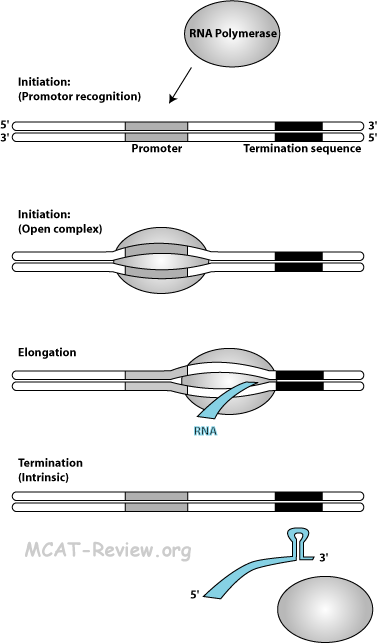
      <div class="fiche-section">
        <div class="fiche-section-title">Traduction</div>
<ul class="fiche-list">- mRNA (messenger RNA): contains codons that code for the peptide sequence.
- tRNA (transfer RNA): contains the anticodon on the "tip" and the corresponding acide aminé on the "tail". Link the correct acide aminé to its corresponding mRNA codon through codon-anticodon interaction.
- rRNA (ribosomal RNA): forms the ribosome. Catalyzes the formation of the liaison peptidique.
- Role and structure of ribosomes
- Ribosome is the enzyme that catalyzes synthèse des protéines.
- Ribosome has 2 subunits - the large and the small.
- The large subunit is responsible for the peptidyl transfer reaction.
- The small subunit is responsible for the recognizing mRNA and binds to the Shine-Dalgarno sequence on the mRNA (Kozak sequence for eukaryotes).
- Both subunits are needed for traduction to occur and they come together in a hamburger fashion that sandwiches the mRNA and tRNAs in between.
- Mechanism of traduction: initiation, termination, co-factors
- Chain Initiation: To begin traduction, you need to form the complexe d'initiation. The complexe d'initiation is basically an assembly of everything needed to begin traduction. This includes mRNA, initiator tRNA (fmet), and the ribosome (initiation factors, and GTP aids in the formation of the complexe d'initiation). The complexe d'initiation forms around the codon d'initiation (AUG), which is just down stream of the Shine-Dalgarno sequence. The Shine-Dalgarno sequence is the "promoter" equivalent of traduction for prokaryotes (Kozak sequence for eukaryotes).
- Chain Elongation: protéine is made from the N terminus to the C terminus. mRNA codons are read from the 5' to the 3' end. Elongation consists of:
- Binding: new tRNA with its acide aminé (tRNA+acide aminé is called aminoacyl-tRNA) enters the A site. GTP and facteur d'élongation required.
- Peptidyl transfer: attachment of the new acide aminé to the existing chain in the P site. The mechanism is a little strange, what happens is that the already existing chain in the P site migrates and attaches to the aminoacyl-tRNA in the A site.
- Translocation: the lone tRNA in the P site gets kicked off (E site), and the tRNA in the A site, along with the peptide chain attached to it, moves into the P site. The mRNA gets dragged along also - the codon that was in the A site is now in the P site after translocation. The A site is now empty and ready for the binding of a new aminoacyl-tRNA to a new codon. Elongation factor and GTP required.
- Chain termination: When a codon stop is encountered, protéines called facteurs de libération, bound to GTP, come in and blocks the A site. The peptide chain gets cleaved from the tRNA in the P site. Peptide chain falls off, and then the whole traduction complex falls apart.
- Amino acid activation: enzymes called aminoacyl-tRNA synthetases attach the correct acides aminés to their corresponding tRNAs. ATP required.
- Post-translational modification of protéines
- Add carbohydrate groups: localization tags, gives mucous texture to proteoglycans
- Adding fatty groups: membrane anchored protéines
- Acétylation: localization tags
- Disulfide bond formation: joins different subunits
- Phosphorylation: activating/inactivating an enzyme
- Ubiquination: tags protéine for destruction
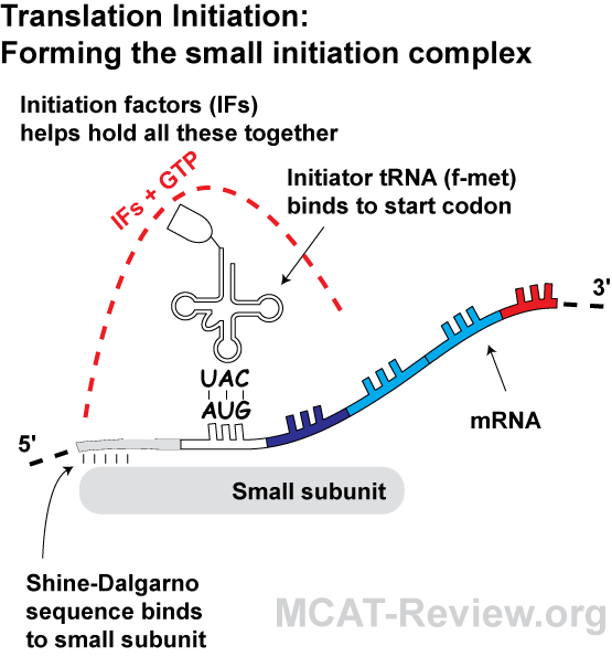
<div class="fiche-section">
<div class="fiche-section-title">Diagramme de la traduction - aperçu graphique de l'initiation, l'élongation et la terminaison</div>
<ul class="fiche-list">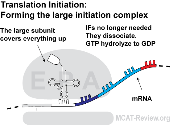
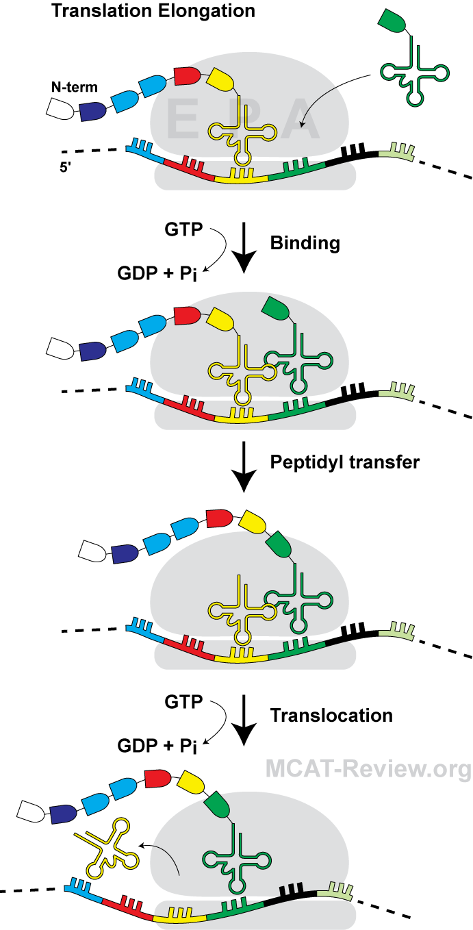
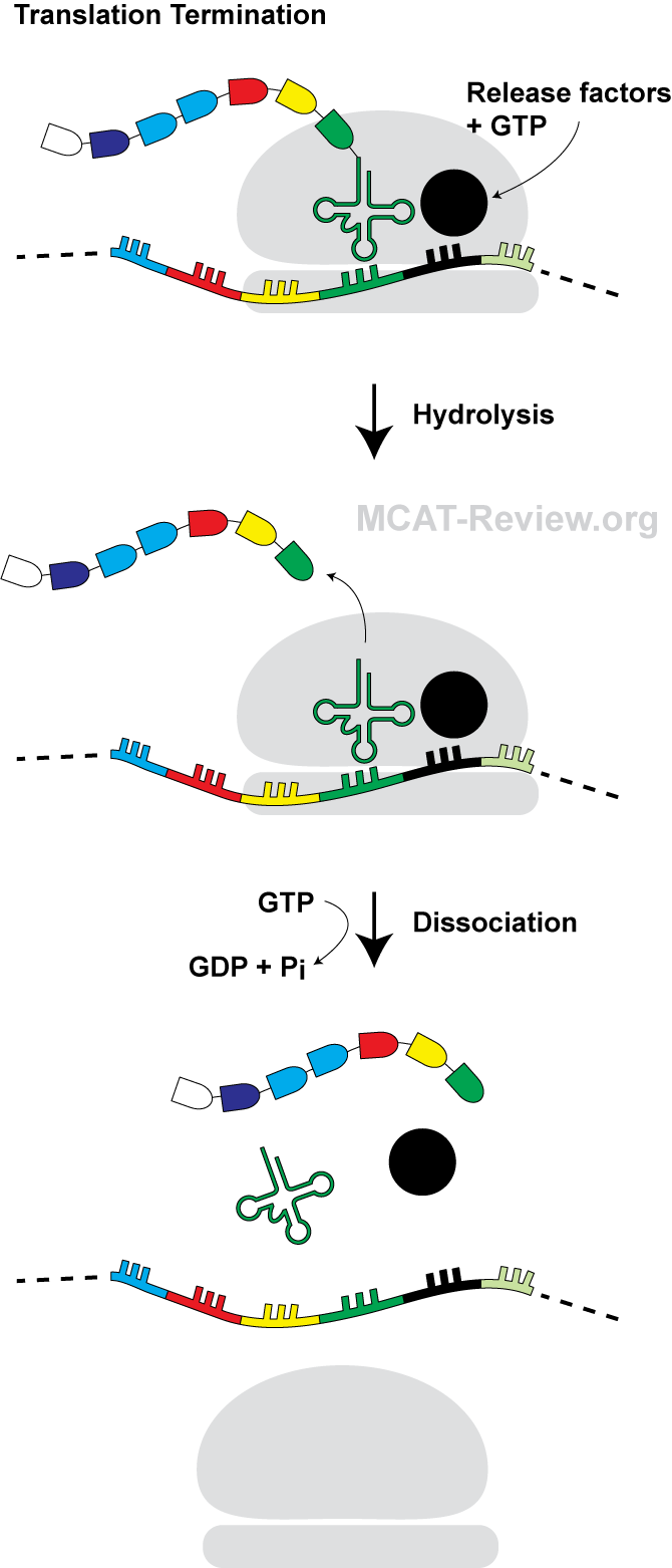
      <div class="source-note">Contenu adapté de <a href="https://mcat-review.org/molecular-biology-protein.php" target="_blank" rel="noopener">mcat-review.org</a> — Traduction et mise en forme par Logopoïos</div>
    </div>
  </div>

  <div class="chapter" id="ch-12">
    <div class="chapter-header" onclick="toggleChapter(this)">
      <div>
        <div class="chapter-num">Chapitre 12</div>
        <div class="chapter-title">Systèmes musculaire et squelettique</div>
      </div>
      <div class="chapter-toggle">▼</div>
    </div>
    <div class="chapter-body">

      <div class="fiche-section">
        <div class="fiche-section-title">Système musculaire</div>
<ul class="fiche-list">- support, mobility
- Support = muscles maintain your posture when you sit/stand, muscles also stabilize joints, help prevent dislocations.
- Mobility = you move because of muscles squelettiques. Your guts move because of muscles lisses. Your flux sanguin because of pumping action of the heart.
- peripheral circulatory assistance
- Heart is a muscle that pumps blood.
- Contraction of muscles squelettiques around the deep veines help squeeze the blood through those veines.
- Diaphragme contraction (breathing) sucks blood into the chest cavity, and also squeezes on abdominal veines.
- thermorégulation (shivering reflex)
- Muscles generate heat when you shiver in response to cold.
- Structural characteristics of skeletal, smooth, and muscle cardiaque; striated vs nonstriated
- Skeletal muscle = striated, voluntary, shaped like long fibers, multinucleated.
- Smooth muscle = nonstriated, involuntary, shaped like almonds (tapered ends), one noyau per cellule.
- Cardiac muscle = striated, involuntary, branched, shaped like fibers cross-linked to one another, typically one noyau per cellule.
- Striated = due to sarcomere structure (A bands dark, I bands light). Skeletal and muscles cardiaques have sarcomeres.
- Nonstriated = muscles lisses don't have sarcomeres so they're not striated. They still have myosin, actin, and use the mécanisme du filament glissant. They just are not organized into sarcomeres.
- Read more about sarcomeres here.
- Nervous control
- neurones moteurs = efferent neurones = signals muscles/organes to do stuff = the opposite of neurones sensoriels.
- Somatic neurones moteurs = controls muscles squelettiques.
- Autonomic neurones moteurs = sympathetic and division parasympathiques = controls involuntary (smooth, cardiac) muscles.
- jonctions neuromusculaires, plaques motrices
- jonction neuromusculaire = nerf (terminaison axonale) meets muscle (plaque motrice).
- plaque motrice = part of muscle membrane cellulaire (sarcolemma) that synapse with the neurone moteur, has récepteurs for the neurotransmetteurs.
- what happens at the jonction neuromusculaire? Action potential of nerf reach terminaison axonale → release neurotransmetteurs into synapse → récepteurs on plaque motrice (sarcolemma) picks this signal → potentiel gradué created → if reaches threshold, then potentiel d'action created → potentiel d'action travels down the sarcolemma and cause muscle to contract.
- voluntary and involuntary muscles
- voluntary = you can control = muscles squelettiques, eg. Biceps.
- involuntary = you can't control = smooth (eg. gut) and cardiac (heart) muscles.
- sympathetic and parasympathetic innervation
- sympathetic = lutte ou fuite = heart beat faster, pupil dilation, raise pression artérielle, blood to muscles, less blood to système digestif.
- parasympathetic = rest and digest = opposite of sympathetic = heart slower, pupil constriction, lower pression artérielle, blood to système digestif.
- Both sympathetic and parasympathetic are neurones moteurs that innervate involuntary muscles.
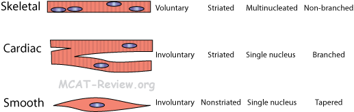
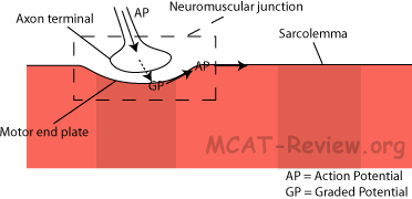
      <div class="fiche-section">
        <div class="fiche-section-title">Système squelettique</div>
<ul class="fiche-list">- structural rigidity and support: bone forms the body's framework.
- calcium storage: bone stores calcium. When calcémie is low, parathyroid hormones signal bone tissu to break down and release calcium.
- physical protection: rib cage protects internal organes. Skull protects cerveau. Spine protects moelle épinière. Many large bones also shelter moelle osseuse that contains cellules souches that make blood.
- Skeletal structure
- spécialisation of bone types, structures
- Long bones: shaped like a rod. eg. arm, leg, finger bones.
- Short bones: shaped like a cube. eg. wrist, ankle bones.
- Flat bones: bones that are flat. eg. sternum, shoulder blades, ribs, skull.
- Irregular bones: complicated shapes. eg. vertebrae, hip.
- joint structures
- Joint = where bone meets bone.
- Joints can be mobile or non-mobile.
- Mobile joints (synovial) have a fluid-containing cavity to lubricate movements of the bones.
- Non-mobile joints connect bone to bone with cartilage or fiber.
- Ball and socket joint: shoulder, hip.
- Hinge joint: elbow.
- Gliding joint: wrist.
- Immobile joint: plates of the skull, rib-to-sternum.
- The joint type that allows most freedom of movement = ball-and-socket.
- endoskeleton versus exoskeleton
- Endoskeleton = what we have, skeleton on the inside.
- Exoskeleton = what insects have, skeleton (chitin) on the outside.
- Cartilage (structure and function)
- Cartilage = cellules + matrice extracellulaire.
- Cartilage cellules = chondrocytes.
- Extracellular matrix = secreted by the cellules, contains fiber meshworks that give the cartilage its characteristic properties (flexibility and resilience)
- Fonctions
- Flexibility: ear, nose, épiglotte, end of ribs
- Resilience, compressibility: Ends of bones in joints, knee, between vertebrae.
- Ligaments, tendons
- Ligament = connect bone to bone, stabilize joints.
- Tendon = connect muscle to bone, anchors muscle.
- Bone structure
- Macroscopically: bone = solid strucuture with canals inside where vaisseau sanguin runs, and holes where cellules can reside, the whole thing surrounded by membrane that contains cellules souches (osteoblasts) and osteoclasts.
- Microscopically: bone = cellule + matrice extracellulaire = arranged in cylinders called osteons, with vaisseau sanguin and nerf running through the middle of the cylinder.
- Cellule = osteocytes (bone cellules).
- calcium-protéine matrix: the matrice extracellulaire of bone consists of calcium salts, collagen fibers, and ground substance (glue).
- bone growth (osteoblasts, osteoclasts)
- Growth in length:
- Lengthwise bone growth occurs at the ends of long bones at the knobs.
- Osteoblasts' role in lengthwise bone growth is to add bone tissu at the bone ends.
- By itself, osteoblasts will lengthen the knobs at the ends of the bone.
- Osteoclasts' role in bone growth is to remodel bone tissu by chipping away the knobs until it's the right size and shape.
- Growth in diameter:
- Osteoblasts' role in diameter growth of bones is to add bone tissu to the outside of the bone.
- Osteoclasts' role in diameter growth of bones is to remove some bone tissu from the inside of the bone (bones are hollow).
- Without osteoclasts, diameter growth will result in bones that are too thick and too heavy. Even with osteoclasts, bones still grow thicker, just not unwieldly thick.
- Osteoblasts vs osteoclasts vs osteocytes
- Osteoblasts = cellules souches that give rise to osteocytes = builds bone.
- Osteocytes = mature bones cellules = reside in bone for housekeeping.
- Osteoclasts = large cellules that break down bone.
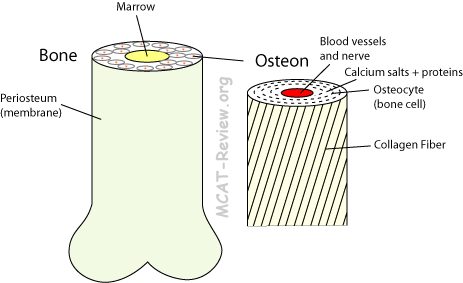
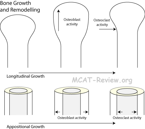
      <div class="source-note">Contenu adapté de <a href="https://mcat-review.org/muscle-skeletal-systems.php" target="_blank" rel="noopener">mcat-review.org</a> — Traduction et mise en forme par Logopoïos</div>
    </div>
  </div>

  <div class="chapter" id="ch-13">
    <div class="chapter-header" onclick="toggleChapter(this)">
      <div>
        <div class="chapter-num">Chapitre 13</div>
        <div class="chapter-title">Systèmes nerveux et endocrinien</div>
      </div>
      <div class="chapter-toggle">▼</div>
    </div>
    <div class="chapter-body">

      <div class="fiche-section">
        <div class="fiche-section-title">Système nerveux : Structure et fonction</div>
<ul class="fiche-list">- high-level control and integration of body systems
- response to external influences
- entrée sensorielle
- sensory = afferent
- influx nerveux conveyed to the CNS.
- sortie motrice
- motor = efferent
- influx nerveux from the CNS to effector organes.
- integrative and cognitive abilities
- Organization of vertebrate système nerveux
- CNS = Central Nervous System = Cerveau and moelle épinière
- Cerveau
- Spinal Cord
- PNS = Peripheral Nervous System = Everything else
- Sensory = Afferent = Nerfs carrying signal toward CNS.
- Motor = Efferent = Nerfs carry signal toward effector organes.
- Somatic Nervous System = Voluntary = Controls muscles squelettiques.
- Autonomic Nervous System = Involuntary = Effects visceral organes.
- Sympathetic division = réponse de lutte ou fuite.
- Paradivision sympathique = Rest.
- Sensor and effector neurones
- Sensor = senses, carries sensory signals from the body to the CNS.
- Effector = causes an effect = carries motor signals from the CNS to the body.
- Sympathetic and parasympathetic système nerveuxs (functions, antagonistic control)
- Sympathetic = prepares body for activity = réponse de lutte ou fuite.
- Increase fréquence cardiaque, pression artérielle
- More flux sanguin to muscles, less to système digestif.
- Pupil dilation.
- Break down glycogen to release glucose into blood.
- Parasympathetic = prepares body to rest
- Decrease fréquence cardiaque, pression artérielle.
- Less blood to muscles, more to système digestif.
- Pupil constriction.
- Synthesizes glycogen for storage from glucose.
- Reflexes
- boucle de rétroaction, arc réflexe, effects on flexor and extensor muscles
- Feedback loop = rétroaction positive (reinforce initial event), rétroaction négative (counteracts initial event), or arc réflexe (usually a type of rétroaction négative).
- rétroaction positive = uterine contraction lead to oxytocin release, which causes more uterine contraction.
- rétroaction positive = coagulation sanguine platelets activated at wound site attract more platelet activation and clumping.
- rétroaction négative = drop in pression artérielle causes ADH release, which increases it. Conversely increase in pression artérielle causes a drop in ADH.
- Reflex arc = withdrawal from a painful stimulus = rétroaction négative.
- Reflex arc = knee jerk = tapping the knee tendon causes sudden stretching of the muscle, which lead to contraction of that muscle that creates the knee jerk = rétroaction négative.
- Reflex arc = récepteur → neurone sensoriel → integration center → neurone moteur → effector
- récepteur = site of stimulus
- neurone sensoriel = carries impulse from récepteur to integration center
- integration center = connects sensory to neurone moteur via synapse inside the CNS
monosynaptic = no interneuron, direct synapse of sensory to motor.
- polysynaptic = interneuron(s) present.
- neurone moteur = carries impulse toward effector.
- effector = site of response to the stimulus
- Examples of reflexes: knee-jerk, withdrawal from pain
- Effects on flexor and extensor muscles
- During the knee-jerk, in addition to contracting the extensor, the reflex relaxes the flexor.
- Golgi tendon reflex: sudden contraction of the quads (extensor), causes a rétroaction négative that relaxes the quads and contracts the hamstrings (flexor).
- role of moelle épinière, cerveau
- Spinal cord provides the synapse (or synapses if it's polysynaptic) for the arc réflexe.
- Even though the arc réflexe bypasses the cerveau, the cerveau is still aware of it happening.
- efferent control
- Cerveau can override spinal reflexes (eg. you don't jerk away from getting a vaccine shot)
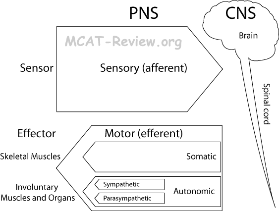
      <div class="fiche-section">
        <div class="fiche-section-title">Système nerveux : Réception et traitement sensoriel</div>
<ul class="fiche-list">- Skin: touch, heat and pain récepteurs close to the surface (dermis-epidermis boundary), pressure récepteurs deeper in the dermis.
- proprioceptor: senses the position of a body part, located in muscle and tissu conjonctif.
- somatic sensors:
- mechanoreceptors - touch, pressure
- thermoreceptor - temperature change (a warm object will feel warm if your hand is cool, but won't feel warm if your hand is already warm)
- photoreceptor - light
- chemoreceptor - taste, smell
- nocioreptors - pain (extreme heat, cold, pressure, chemicals)
- Olfaction, taste
- Olfaction:
- Chemicals enter the nose via nostrils.
- Gets into the nasal cavity.
- Trapped in the mucus on top of the nasal cavity.
- Picked up by the membrane récepteurs on cilia (non-mobile, but they increase the surface) of the olfactory récepteur cellule.
- Causes cellule dépolarisation, and subsequent transduction of signal to the cerveau.
- Taste:
- Chemicals dissolve in saliva.
- Carried inside taste bud
- Hair-like microvilli of taste cellules inside taste bud picks up chemicals.
- Releases neurotransmetteurs to send signal to cerveau.
- Hearing
- ear structure
- Ear canal = auditory canal.
- Tympanic membrane = eardrum.
- Ear bones = malleus (hammer) → incus (anvil) → stapes (stirrup).
- Vestibule = contacts the oval window (where stirrup vibrates), is continuous with semicircular canals and cochlea.
- Cochlea = spiral = houses hair cellules.
- Semicircular canals = 3 of them perpendicular to one another = senses position and movement of the head, help you balance.
- mechanism of hearing
- Sound enters ear.
- Hits ear drum (tympanic membrane)
- Malleus (hammer) → Incus (anvil) → Stapes (stirrup)
- Vibrates fluid in Cochlea.
- Transmits to fluid in Cochlea.
- Cochlear hair cellules excited by vibrations, and sends signal to cerveau.
- Vision
- light récepteurs
- Photoreceptor cellules located on the back of the retina.
- Rods = senses light and dark (no color), more sensitive.
- Cones = senses color, less sensitive.
- Rhodopsin = chemical responsible for light reception = Retinal (chemical) + Opsin (transmembrane protéine)
- Light converts cis-retinal → trans-retinal.
- trans-retinal then causes hyperpolarisation of photoreceptor cellule, which prompts the chain of events that sends signal to the cerveau.
- Sends signal to cerveau via a bundle of nerfs on the back of the retina (where the blind spot is)
- eye structure
- Light first travels through the cornea
- Through the pupil (hole in the iris muscle)
- Lens = focuses light on retina.
- Vitreous humor = fluid.
- Retina = screen on the back of the eye = contains photoreceptors.
- visual image processing
- The lens of the eye, just like a convex lens in physics, forms a real image on the retina.
- Real images are inverted.
- The cerveau processes this inverted image to make it seem upright in your mind.
- The cerveau combines the two images from each eye to make a 3D image, from which you can judge distance.
- Another reason for combining the two images from both eyes is that it gets rid of the blind spot in each eye.
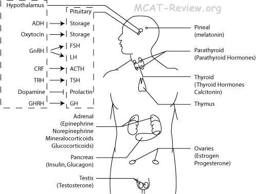
      <div class="fiche-section">
        <div class="fiche-section-title">Système endocrinien : Hormones</div>
<ul class="fiche-list">- Endocrine system = make hormones = specific control of all cellules cibles of that hormone.
- Definition of glande endocrine, hormone
- endo = within, crine = to secrete
- glandes endocrines secreting hormones into surrounding tissu fluids.
- endocrine vs. exocrine, autocrine, paracrine
- endocrine: hormone, no duct, acts long distances
- exocrine: non-hormone secretions into ducts.
- autocrine: local chemicals, act short distances on themselves
- paracrine: local chemicals, act short distances on other cellules
- hormone = chemicals that regulate métabolisme and function of cellules.
- Major glandes endocrines (names, locations, produits)
- Hypothalamus: Releasing hormones for the pituitary, ADH and oxytocin.
- Releasing hormones/factors stimulates pituitary to release its hormone.
- GnRH = Gonadotropin Releasing Hormone = stimulates pituitary to release FSH and LH.
- CRF = Corticotropin Releasing Factor.
- TRH = Thyroid Releasing Hormone.
- Dopamine = inhibits prolactin release.
- GHRH = Growth Hormone Releasing Hormone.
- ADH = Antidiuretic Hormone = Vasopressin = increase réabsorption d'eau in rein = conserve water, increase pression artérielle.
- Oxytocin = stimulates uterine contractions during labor, also milk sécrétion during suckling.
- Pituitary: makes FLAT PEG, stores ADH and oxytocin.
- FSH = Follicle Stimulating Hormone = Stimulate ovaire follicles to mature, testicule to produce spermatozoïde.
- LH = Luteinizing Hormone = LH surge triggers ovulation, stimulates testicule to produce testosterone.
- ACTH = AdrenoCorticoTropic Hormone = Stimulates adrenal cortex to release glucocorticoids and mineralocorticoids.
- TSH = Thyroid Stimulation Hormone = Stimulate thyroid to release thyroid hormones.
- PRL = Prolactin = Stimulates breast to produce milk.
- E = Endorphins.
- GH = Growth Hormone = Stimulates growth of muscle, bone, burns fat.
- Pineal: makes melatonin, which makes you sleepy at night.
- Thyroid
- Thyroid hormones: increase métabolisme, requires iodine.
- Calcitonin: turns blood Ca2+ into bone. Lowers blood Ca2+.
- Parathyroid: makes Parathyroid Hormone (PTH), which increases blood Ca2+ by bone resorption, dietary calcium absorption, and calcium reabsorption in reins.
- Thymus: Thymus hormones (thymo-, thymic), stimulates cellules T to develop.
- Adrenal
- Epinephrine and norepinephrine = réponse de lutte ou fuite
- Mineralocorticoids = aldosterone = increase Na+ and water retention, raises pression artérielle.
- Glucocorticoids = cortisol = stress hormone = increase glycémie.
- Androgens = testosterone.
- Pancréas
- Glucagon = increases glycémie (break down glycogen, stimulate néoglucogenèse).
- Insulin = lower glycémie (stimulates glucose uptake by cellules).
- Ovaire: make estrogen (and a small amount of testosterone).
- testicule: make testosterone.
- Endocrine diseases
- Diabetes
- no insulin made, or no insulin récepteurs
- glucose can't enter cellules
- high glycémie
- cellule starved of sugar, leading to acide gras métabolisme, which leads to production of ketone bodies, which lead to ketoacidosis (more acidic blood).
- sugar in urine, leading to more water in urine due to osmose.
- Hypothyroidism
- Decreased thyroid hormone.
- Low métabolisme.
- If cause of disease is lack of iodine in diet, then goiter develops from an accumulation of thyroid hormone precursor lacking iodine.
- Hyperthyroidism
- Too much thyroid hormone.
- High métabolisme.
- Gigantism = too much Growth Hormone during growing age = well-proportioned giants.
- Acromegaly = too much Growth Hormone later on in life = disproportioned growth of certain areas of the body (the parts that still respond to growth hormone).
- Major types of hormones
- acide aminé based = acide aminé derivatives = most hormones are this type.
- steroids = cholesterol derivatives = testosterone, estrogen, adrenocortical hormones.
</div>
<div class="fiche-section">
<div class="fiche-section-title">Système endocrinien : Mécanismes d'action des hormones</div>
<ul class="fiche-list">- water soluble hormones
- Can't cross the membrane plasmique.
- Bind to membrane récepteurs on the outside of cellules.
- Secondary messengers then relay the signal inside the cellule.
- lipid-soluble hormones
- Able to cross the membrane plasmique.
- Directly activate gènes.
- cAMP pathway:
- Amino acid hormone binds membrane récepteur.
- G protéine activated.
- Adenylate cyclase activated.
- cAMP made.
- Protéine kinase cascade.
- Phospholipid pathway:
- Amino acid hormone binds membrane récepteur.
- G protéine activated.
- Phospholipase C activated.
- Membrane phospholipid split into DAG and IP3.
- DAG triggers protéine kinase cascade.
- IP3 releases Ca2+ from the ER.
- Steroid pathway:
- Steroid hormone (and thyroid hormone even though it's acide aminé based) goes inside the cellule.
- Hormone binds récepteur inside the cellule (cytoplasme or noyau).
- Hormone-récepteur complex (facteur de transcription) turns certain gènes on inside the noyau.
- Transport of hormones (flux sanguin): hormones travel long distances via blood and lymph.
- Specificity of hormones (target tissu)
- Specificity depends on the cellules cibles having the récepteurs for the hormone, and non-cellules cibles lacking récepteurs for the hormone.
- Cellules can either upregulate or downregulate the récepteurs they express.
- Integration with système nerveux (feedback control)
- The système nerveux can modulate and override normal control of hormones based on the status of the body. For example, the body's blood "normal" glucose level is set higher when you're under stress.
- Hormones can modulate the système nerveux. For example, low estrogen levels during menses give you a bad mood.
- Normal control of hormones
- Humoral: glands directly respond to chemical levels in the blood (parathyroid respond to low calcémie).
- Neural: glands release hormones when stimulated by nerfs (réponse de lutte ou fuite).
- Hormonal: glands release hormones when stimulated by other hormones (tropic hormones).
      </div>

      <div class="source-note">Contenu adapté de <a href="https://mcat-review.org/nervous-endocrine-systems.php" target="_blank" rel="noopener">mcat-review.org</a> — Traduction et mise en forme par Logopoïos</div>
    </div>
  </div>

  <div class="chapter" id="ch-14">
    <div class="chapter-header" onclick="toggleChapter(this)">
      <div>
        <div class="chapter-num">Chapitre 14</div>
        <div class="chapter-title">Système reproducteur et développement</div>
      </div>
      <div class="chapter-toggle">▼</div>
    </div>
    <div class="chapter-body">

      <div class="fiche-section">
        <div class="fiche-section-title">Système reproducteur</div>
<ul class="fiche-list">- gonads
- male: testicules
- makes spermatozoïde in the tubes séminifères.
- makes testosterone.
- external.
- female: ovaires
- houses immature ovule, which matures monthly after puberty.
- makes estrogen.
- internal.
- genitalia
- male: testicules, penis, and various ducts and glands.
- spermatozoïde made in the tubes séminifères.
- stored in the epididymis.
- travels through vas deferens → ejaculatory duct → urètre → penis
- mnemonic: seven up = Seminiferous tubules, Epididymis, Vas deferens, Ejaculatory duct, nothing, Urètre, Penis.
- female: ovaires, trompes de Fallope, utérus, vagina
- Monthly cycle: ovocyte primaire matures into ovocyte secondaire every month. To prepare for it, the endometrium thickens. If fécondation doesn't occur, menses occur, and the cycle begins anew.
- GnRH = stimulates release of FSH and LH.
- FSH = folicle stimulating hormone = stimulates growth and maturation of follicle.
- Follicle = houses oocyte and produces estrogen.
- Estrogen = normally inhibits LH and FSH, but causes LH surge when it reaches a certain threshold.
- Estrogen reaches this threshold → surge of LH occurs.
- LH = leutinizing hormone = luteinizing hormone = stimulates the outer cellules of the follicle = turns it into corps jaune + maintains it.
- LH surge triggers ovocyte primaire → ovocyte secondaire → rupture of follicle.
- Corpus luteum = makes estrogen and progesterone = maintains endometrium.
- No fécondation → LH falls → corps jaune dies → estrogen and progesterone fall → endometrium dies (menses) → cycle begins anew with FSH and LH re-rising.
- Fécondation occurs → implanted embryon releases hCG → hCG mimics LH to maintain corps jaune → estrogen and progesterone maintained by corps jaune → placenta takes over the responsibility of making estrogen and progesterone later on.
- differences between male and female structures
- male: mostly external. Shared passage with urinary tract.
- female: mostly internal. Separate passage from urinary tract.
- Gametogenesis by méiose
- Male = spermatogenesis = occurs in the tubes séminifères.
- Spermatogonium (2n) = cellule souche. Mitose of spermatogonium can either create more spermatogonium or create spermatocyte primaire.
- Spermatogonium (2n) → mitose → spermatocyte primaire (2n). Occurs after puberty.
- Primary spermatocyte (2n) → méiose I → Secondary spermatocyte (n).
- Secondary spermatocyte (n) → méiose II → spermatid (n).
- Spermatid (n) → mature → spermatozoïde (n). The fancy name for spermatozoïde is spermatozoa.
- Female = oogenesis = occurs in the ovaires, then trompes de Fallope.
- Oogonium (2n) = cellule souche.
- Oogonium (2n) → mitose → ovocyte primaire.
- Primary oocyte (2n) arrests at prophase I (occurs before birth). One comes out of arrest every month (between puberty and menopause).
- Primary oocyte (2n) → méiose I → ovocyte secondaire (n). Ruptures from ovaire follicle into the trompe de Fallope.
- Secondary oocyte (n) arrests at métaphase II. Comes out of arrest if fécondation occurs.
- Secondary oocyte (n) → méiose II → ovum (n).
- Ovum and spermatozoïde
- differences in formation
Male and female gametogenesis side by side
Male
Female
Difference
Spermatogonium (2n)
Oogonium (2n)
Spermatogonium renews its population by mitose throughout life. Oogonium stops renewing its population sometime before birth
Primary spermatocyte
Primary oocyte
Primary oocye arrests at prophase I
Secondary spermatocyte
Secondary oocyte
Secondary oocyte arrests at métaphase II
Spermatozoïde
Ovum
Between the spermatocyte secondaire and the spermatozoïde, there's the spermatid
- differences in morphology
- Spermatozoïde = motile = flagella.
- Ovule = non-motile = round.
- relative contribution to next generation
- Spermatozoïde contributes DNA only (the ovule actively destroys spermatozoïde mitochondries).
- Ovule contributes DNA + everything else (mitochondries, organites, epigenetics).
- Reproductive sequence (fécondation, implantation, development, birth)
- fécondation: spermatozoïde + ovule → zygote
- implantation:
- zygote
- morula (solid ball)
- blastula (sea urchins) or blastocyst (mammals)
- the blastocyst is the one that implants in the endometrium
- development:
- zygote
- blastocyst
- implantation
- gastrulation
- organogenèse
- Birth:
- Switch from getting oxygen from mom's blood → breathing.
- Switch from getting nutriments from mom's blood → suckling.
- Fetal circulation (which bypasses poumons and foie) → normal circulation (by closing off ducts and opennings).
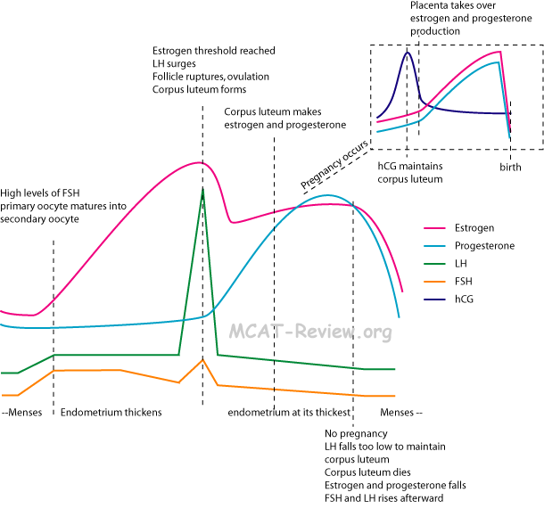
      <div class="fiche-section">
        <div class="fiche-section-title">Embryogenèse</div>
<ul class="fiche-list">- fécondation
- Spermatozoïde meets ovule
- Acrosomal reaction causes spermatozoïde to penetrate ovule
- Cortical reaction causes ovule to prevent additional spermatozoïde from penetrating
- Ovule completes méiose II
- Spermatozoïde and ovule nuclei fuse
- cleavage
- Normal mitotic division cellulaires: cellule grows then divides, grows again, then divides.
- Cleavage = division mitotiques without croissance cellulaire.
- blastula formation
- fécondation produces zygote
- cleavage produces a solid ball called the morula
- morula hollows out into the blastula or blastocyst
- blastula occurs in non-mammals
- blastocyst occurs in mammals
- blastocyst implants
- gastrulation
- first cellule movements
- Cellules from the surface migrate inwards.
- gastrulation occurs slightly different for different animals. Some by invagination, some by migration, some by splitting.
- In mammals, the cellules start migrating inward at the ligne primitive.
- formation of primary germlayers (endoderm, mesoderm, ectoderm)
- The cellules that migrate inwards form the endoderm.
- The cellules that remain outside is the ectoderm.
- The cellules in the middle are the mesoderm.
- neurulation
- ectoderm → cerveau and moelle épinière
- the ectoderm does so by folding into a tube
- Major structures arising out of primary feuillets embryonnaires
- endoderm = innermost layer = guts, poumons, and digestive internal organes (foie, pancréas).
- mesoderm = middle layer = muscle, blood and bone tissus, and interal organes (rein and gonads).
- ectoderm = outermost layer = skin and nerfs (including the cerveau).
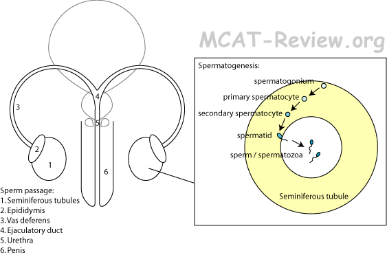
      <div class="fiche-section">
        <div class="fiche-section-title">Mécanismes du développement</div>
<ul class="fiche-list">- commitment = specification followed by determination
- specification = cellule is just beginning to be commited to develope into a certain type cellulaire. The commitment can be reversed at this stage.
- determination = irreversible commitment to become a certain type cellulaire.
- différenciation = becoming a type cellulaire and adopting its specialized functions.
- epidermal cellules produce keratin to protect skin against abrasion.
- myocyte produce actin and myosin to make muscles contract.
- neurones make neurotransmetteurs to transmit electrochemical impulses.
- tissu types
- Epithelial: skin, lining of organes
- Connective: blood, bone, tendons, ligaments, cartilage
- Nervous: cerveau, moelle épinière, nerfs
- Muscle: skeletal, smooth, and muscle cardiaque
- Cellule communication in development
- Induction: one group of cellules changing the behavior of an adjacent group of cellules.
- inducer = the one that sends the signal for the other to change.
- responder = the one that gets the signal and changes.
- For example, the optic vesicle is able to induce the ectoderm to develope into lens.
- Another example is the induction of wing feathers in the chick by the dermal mesenchyme.
- Induction mechanisms: physical touching of cellules (juxtracine) or by releasing chemicals (paracrine).
- Cellule migration
- Gastrulation, crête neurale migration cellulaire
- Hirschprungs disease: defective crête neurale migration cellulaire
- Pluripotency: cellules souches (able to differentiate into other types cellulaires)
- Stem cellules in bone can differentiate into osteoblasts or osteocytes
- Stem cellules in moelle osseuse can differentiate into neutrophils, lymphocytes, globules rouges or platelets
- Gène régulation in development
- Differential gène transcription:
- modification of DNA (methylations) can shut off or turn on gènes.
- modification on histones (methylations, acetylations) that wrap the DNA can shut off or turn on gènes.
- to make or not to make facteurs de transcription can regulate what gènes get transcribed.
- Differential maturation de l'ARN:
- selecting what RNA make it outside the noyau to be translated.
- alternative splicing of RNA.
- Traduction régulation
- some mRNA are made to last longer than others (more protéines translated off of it), and some are made to be rapidly degraded (less protéines translated off of it).
- selective inhibition of traduction of stored RNA in the oocyte. Get translated only when needed after fécondation.
- Post-translational régulation
- some protéines are inactive until modified by certain enzymes.
- active protéines can be selectively marked for dégradation by ubiquitin.
- Programmed cellule death
- apoptose = mort cellulaire programmée.
- During apoptose, strong proteases are activated and they digest the cellule from within. In mammals, the proteases are called caspases.
- The spaces between our fingers are created by apoptose.
- The tail of a tadpole undergoes apoptose when it morphs into a frog.
- Existence of regenerative capacity in various species
- Newts can regenerate limbs
- Cellules in their limb stump can dedifferentiate and revert back to cellules souches
- Senescence and aging
- Every time a cellule replicates, the telomere shortens. Eventually, they run out of telomeres
- As a person age, their cellules acquire irreversible dommages à l'ADN by rayonnement and chemicals. Not all can be repaired, and they accumulate
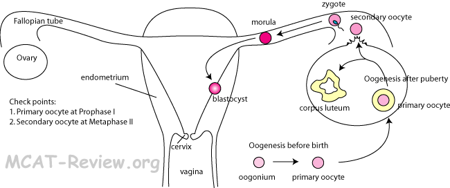
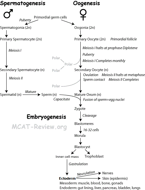
      <div class="source-note">Contenu adapté de <a href="https://mcat-review.org/reproductive-system-development.php" target="_blank" rel="noopener">mcat-review.org</a> — Traduction et mise en forme par Logopoïos</div>
    </div>
  </div>

  <div class="chapter" id="ch-15">
    <div class="chapter-header" onclick="toggleChapter(this)">
      <div>
        <div class="chapter-num">Chapitre 15</div>
        <div class="chapter-title">Système respiratoire</div>
      </div>
      <div class="chapter-toggle">▼</div>
    </div>
    <div class="chapter-body">

      <div class="fiche-section">
        <div class="fiche-section-title">Structure et fonction générales</div>
<ul class="fiche-list">- In poumons: oxygen diffuses into blood. Carbon dioxide diffuses out of blood.
- The mechanism of this échange gazeux follows Henry's law, which basically says that there is an equilibrium concentration of oxygen that should be dissolved in blood.
- When blood reaches the poumons, it has less than the equilibrium concentration of oxygen because the body used the oxygen up. Therefore, oxygen diffuses into blood.
- The CO2 in blood that reaches the poumons is higher than the equilibrium concentration because of the body releases them. Therefore, CO2 diffuses out of blood.
- Thermorégulation: breathing causes you to lose heat (you breathe out warm, moist air).
- protection against disease, particulate matter
- Nostril hair filters out particles.
- Mucus lining of respiratory tract traps agents pathogènes and particles.
- Cilia on mucus lining of respiratory tract sweeps agent pathogène and particles out, where you either spit it out or swallow it into acide gastrique.
- Macrophages reside in alvéoles.
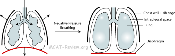
      <div class="fiche-section">
        <div class="fiche-section-title">Mécanismes de la respiration</div>
<ul class="fiche-list">- Diaphragme = muscle that pulls downward when contracting, which increases chest volume, decreases pressure, and sucks air into poumons.
- Rib cage = expands outward during breath intake. Intercostal muscles help this expansion. At rest, the rib cage maintains poumon volume, prevent poumon from collapsing, forms a cage around poumons for protection.
- Differential pressure = difference between intrapulmonary (inside poumon) pressure and intrapleural (outside poumon) pressure.
- Intrapulmonary pressure = pression atmosphérique (poumon is open to the outside, so has same pressure as outside).
- Intrapleural pressure = less than pression atmosphérique = sucks on the poumons, prevent poumon from collapsing. During breath intake, intrapleural pressure decreases even further, causing the poumon to expand.
- Negative pressure mechanism in breathing is just a fancy term for sucking. You breathe in by establishing pression négative in the poumon (sucking). However, when someone gives you mouth-to-mouth, that's pression positive.
- resiliency and tension de surface effects
- Poumon is elastic, it recoils as soon as you relax after breath intake. If not for the rib cage, the poumon would collapse even further.
- Surface tension causes the poumon to collapse. Surfactants produced in the alvéoles decreases tension de surface, and helps alvéoles to stay open.
      </div>

      <div class="source-note">Contenu adapté de <a href="https://mcat-review.org/respiratory-system.php" target="_blank" rel="noopener">mcat-review.org</a> — Traduction et mise en forme par Logopoïos</div>
    </div>
  </div>

  <div class="chapter" id="ch-16">
    <div class="chapter-header" onclick="toggleChapter(this)">
      <div>
        <div class="chapter-num">Chapitre 16</div>
        <div class="chapter-title">Phases et équilibres de phases</div>
      </div>
      <div class="chapter-toggle">▼</div>
    </div>
    <div class="chapter-body">

      <div class="fiche-section">
        <div class="fiche-section-title">Phase gazeuse</div>

        <figure class="fiche-figure">
      <div class="fiche-section">
        <div class="fiche-section-title">Température absolue, échelle K</div>
        <ul class="fiche-list">
K
°C
°F
Absolute zero
0
-273
-460
Freezing point of water / point de fusion of ice
273
0
32
Room temperature
298
25
77
Body temperature
310
37
99
Boiling point of water / condensation of steam
373
100
212
- K = °C + 273
- F = °C x 1.8 + 32
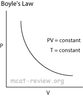
      <div class="fiche-section">
        <div class="fiche-section-title">Pression, baromètre à mercure simple</div>
<ul class="fiche-list">- Due to gravity, the atmosphere exerts a pressure of 101 kPa at sea level. For convenience, 101 kPa = 1 atm.
- Pressure decreases at higher elevations.
- The mercury barometer measures pression atmosphérique by allowing the pression atmosphérique to "push" on a column of mercury.
- The barometer is open at one end and closed off (vacuum) at the other.
- The atmosphere "pushes" at the open end, which results in the mercury rising up in the closed end.
- The measured pression atmosphérique P = F/A. F is the weight of the mercury that got pushed up and A is the cross-section area of the column that the mercury got pushed through.
- Standard mercury barometers are calibrated such that 1 atm of pressure will push the mercury up by 760 mm. For convenience, mm Hg is also called the Torr. So, you don't have to do the P=F/A calculation to find out the pressure reading from a barometer. Just know that 1 atm = 760 mm Hg = 760 torr.
- 1 atm = 101 kPa = 101,000 Pa = 760 mm Hg = 760 Torr.
- When performing P = F/A calculations, make sure that F is in Newtons, A is in meter squared and the resulting P will be in Pascals. You can then convert the Pascals to whatever units the answer choices are in.
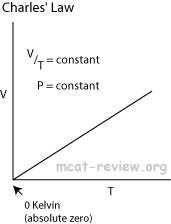
      <div class="fiche-section">
        <div class="fiche-section-title">Volume molaire à 0 degré Celsius et 1 atm = 22,4 L/mol</div>
<ul class="fiche-list">- Another way you can remember this is to look at the periodic table: Air is made up mostly of nitrogen, which has an atomic mass of 14. In the diatomic form, N2 weighs 14x2 = 28 grams per mol. Now, air is really light. In order for you to grab 28 grams of air, you need more than just a bottle of air, you need a huge tank totaling 22.4 L.

      <div class="fiche-section">
        <div class="fiche-section-title">Gaz parfait</div>
<ul class="fiche-list">- An gaz parfait consists of pointy dots moving about randomly and colliding with one another and with the container wall. The gaz parfait obeys the kinetic molecular theory of gases and has the following properties.
- Random molecular motion.
- No forces intermoléculaires.
- No (negligible) molecular volume.
- Perfectly elastic collisions (conservation of total énergie cinétique).
- You can treat gases as gaz parfaits at:
- Low pressures
- High temperatures
- Deviation from the ideal occurs at high pressure and low temperature. At these conditions, the molécules de gaz are "squished" together. When the molécules de gaz are so close together, they experience interactions intermoléculaires. Also, the molecular volume becomes significant when the total volume is squished down so much. The attractions intermoléculaires will cause collisions to be sticky and inelastic. At the extremely high pressures and low temperatures, gases cease to be gases at all - they condense into liquids.
- Ideal gases behave according to the gaz parfait law.
- gaz parfait law PV=nRT, where P is pressure, V is volume, n is # mols of gas, R is the constante des gaz, and T is temperature.
- Combined loi des gaz:
- Because nR is constant (n is the # mols and R is the constante des gaz), PV/T must also be constant.
- Boyle's law and Charles' law can all be derived from the combined loi des gaz.
- Boyle's law: at constant temperature, P1V1 = P2V2
- Charles' law: at constant pressure,
- Charle's law extrapolates to absolute zero, where volume also goes to zero (this is only an extrapolation).
- Avogadro's law: Equal volumes of two gases will also contain equal number of mols of each gas (given ideal conditions: gaz parfait at STP).
- PV = nRT
- R is constant, and at STP, pressure and temperature is also constant.
- V/n = RT/P
- If you plug in STP values, you'll end up with V/n = 22.4 L/mol.
- All gaz parfaits at STP will occupy 22.4 L per mol of molécules de gaz.
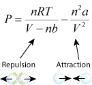
      <div class="fiche-section">
        <div class="fiche-section-title">Théorie cinétique moléculaire des gaz</div>
<ul class="fiche-list">- Random molecular motion.
- No forces intermoléculaires.
- No (negligible) molecular volume.
- Perfectly elastic collisions (conservation of total énergie cinétique).
- The kinetic theory holds the following concepts:
- Pressure of a gas is due to its molecules constantly colliding with the walls of its container.
- Pressure is equally distributed over the walls of the container because molecular motion is random.
- Temperature is a measure of the average énergie cinétique of the molécules de gaz.
- Higher temperature means the molecules are traveling faster, lower temperatures means slower molecules.
- Diffusion and Effusion
- Diffusion: random molecular motion, causing a substance to move from an area of higher concentration to an area of lower concentration (diffusion down its gradient de concentration).
- Effusion: random molecular motion, causing a substance to escape a container through a very small openning.
- Graham's Law (applies both to diffusion and effusion for the purposes of the MCAT).
- Rate1/Rate2 = √M2/M1
- Rate = rate of diffusion or effusion. M = molecular weight of molécule de gaz.
- A possible question on the MCAT is two gasses diffuse down a tube from opposite ends. Where will the gases meet? The gist of this is that the lighter gas will travel faster, and the gases will meet at a point that is farther from the end of the lighter gas.
- Graham's Law is derived from the Kinetic theory
- Temperature = average énergie cinétique
- At a given temperature all gases have the same énergie cinétique.
- ½m1v12 = ½m2v22
- m1v12 = m2v22
- v12/v22 = m2/m1
- v1/v2 = √m2/m1
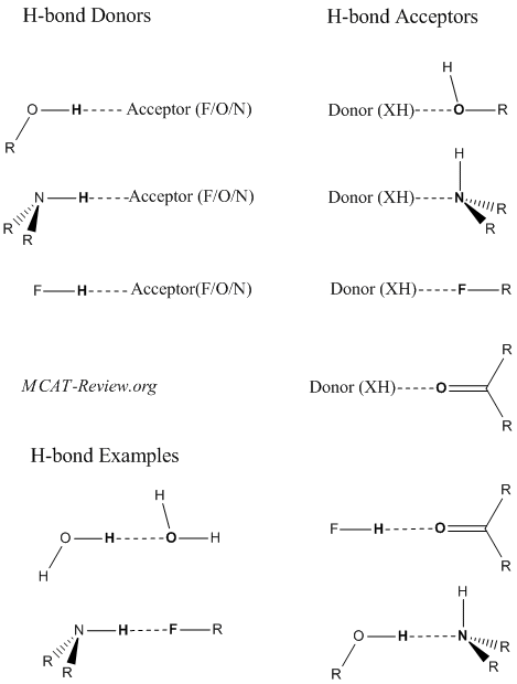
      <div class="fiche-section">
        <div class="fiche-section-title">Déviation du comportement des gaz réels par rapport à la loi des gaz parfaits</div>
<ul class="fiche-list">- When molecules are far apart (under conditions of low P, high T), they are ideal.
- When molecules are brought close together (higher P, lower T), they experience attraction intermoléculaire.
- When molecules are brought so close together that they clash into one another, they experience steric repulsion.
- quantitative (Van der Waals' equation)
- b for bounce. The term with the constant b is the repulsion term. The greater b is, the more repulsion, which leads to greater pressure.
- a for attraction. The term with the constant a is the attraction term. The greater a is, the more attraction, which leads to less pressure.
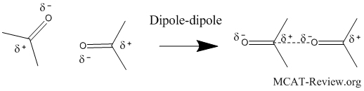
      <div class="fiche-section">
        <div class="fiche-section-title">Pression partielle, fraction molaire</div>
        <ul class="fiche-list">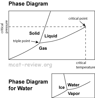
      <div class="fiche-section">
        <div class="fiche-section-title">Loi de Dalton reliant la pression partielle à la composition</div>
<ul class="fiche-list"> </div>
<div class="fiche-section">
<div class="fiche-section-title">Force</div>
</div>
<div class="fiche-section">
<div class="fiche-section-title">Liaison hydrogène</div>
<ul class="fiche-list"> </div>
<div class="fiche-section">
<div class="fiche-section-title">Interactions dipolaires</div>
<ul class="fiche-list"> </div>
<div class="fiche-section">
<div class="fiche-section-title">Force</div>
<ul class="fiche-list"> </div>
<div class="fiche-section">
<div class="fiche-section-title">Équilibres de phases</div>
</div>
<div class="fiche-section">
<div class="fiche-section-title">Changements de phase et diagrammes de phases</div>
<ul class="fiche-list">- Water diagramme de phases is different from others because the solid-liquid boundary is slanted to the left. This is because water (liquid) is more dense than ice (solid), and if you increase the pressure at a given temperature, then you turn ice into water.
- Mnemonic for remembering which section of the diagramme de phases is for gases: "gas comes out this way."
</div>
<div class="fiche-section">
<div class="fiche-section-title">Point de congélation, point de fusion, point d'ébullition, point de condensation</div>
<ul class="fiche-list"> </div>
<div class="fiche-section">
<div class="fiche-section-title">Molalité</div>
<ul class="fiche-list"> </div>
<div class="fiche-section">
<div class="fiche-section-title">Propriétés colligatives</div>
<ul class="fiche-list">- Solute particles in solution like to keep the solution in liquid phase. This is why it makes it harder to boil (raises its point d'ébullition) and also makes it harder to freeze (lowers the point de congélation). Lowering the pression de vapeur is just another fancy name for raising the point d'ébullition.
- Van't Hoff Factor (i): all propriétés colligatives take into consideration of the Van't Hoff factor. Basically, it means convert concentration to reflect the total number of particles in solution. For example, glucose has i of 1 because it doesn't break up in solution. NaCl has i of 2, because in solution, it breaks up into 2 particles Na+ and Cl-.
- pression de vapeur lowering (Raoult's law)
- P = χsolvent·P°solvent
- ΔP = χsolute·P°solvent
- P is the pression de vapeur.
- ΔP is the decrease in pression de vapeur.
- χsolute = mol fraction of the solute = # mols of solute / # total mols of both solute and solvent
- χsolvent = mol fraction of the solvent = # mols of solvent / # total mols of both solute and solvent
- P°solvent is the pression de vapeur of the pure solvent alone.
- When you are calculating χsolute, make sure you take into account of van't Hoff. ie. 1 mols of NaCl in solution is actually 2 mols of particles.
- point d'ébullition elevation (deltaTb = kb*m *i)
- ΔTb = kb·m·i
- ΔTb is the increase in point d'ébullition.
- kb is the molal point d'ébullition constant (like almost every other constants, the MCAT will give it to you).
- m is the molality (mol solute/kg solvent).
- i is van't Hoff factor.
- point de congélation depression (deltaTf = -kf*m *i)
- ΔTf = -kf·m·i
- ΔTf is the decrease in point de congélation (the negative sign shows that the change is a decrease).
- kf is the molal point de congélation constant.
- m is the molality (mol solute/kg solvent).
- i is van't Hoff factor.
- pression osmotique
- π = MRT *i
- π is the pression osmotique.
- M is the molarity in mol/L.
- R is ideal constante des gaz.
- T is the temperature in K.
- Osmotic pressure determines whether and in what direction osmose will occur.
- Osmose is the movement of solvent across a semi-permeable membrane from an area of low solute concentration (high solvent concentration) to an area of high solute concentration (low solvent concentration).
- Solvent will move from an area with low π value to an area with high π value.
      </div>

      <div class="fiche-section">
        <div class="fiche-section-title">Colloïdes</div>
<ul class="fiche-list"> </div>
<div class="fiche-section">
<div class="fiche-section-title">Loi de Henry</div>
<ul class="fiche-list"> </div>
<div class="source-note">Contenu adapté de <a href="https://mcat-review.org/phases-equilibria.php" target="_blank" rel="noopener">mcat-review.org</a> — Traduction et mise en forme par Logopoïos</div>
</div>
</div>
<div class="chapter" id="ch-17">
<div class="chapter-header" onclick="toggleChapter(this)">
<div>
<div class="chapter-num">Chapitre 17</div>
<div class="chapter-title">Système tégumentaire</div>
</div>
<div class="chapter-toggle">▼</div>
</div>
<div class="chapter-body">
<div class="fiche-section">
<div class="fiche-section-title">Fonctions dans l'homéostasie et l'osmorégulation</div>
<ul class="fiche-list">- Too cold: hair stands up (goose bumps), vasoconstriction decreases blood supply at skin (less heat loss).
- Too hot: sweat (evaporative cooling), vasodilatation increases blood supply at skin (more heat loss).
- Water homéostasie: Insulates body against water loss.
- Osmorégulation: sweat excretes salts and nitrogenous wastes (urea, acide urique, ammonia)
- Some other functions of the skin:
- protect against UV rayonnement by making melanin (absorbs UV)
- make vitamin D upon exposure to sunlight.
- Act as blood reservoir. Vasoconstriction in skin shunts blood to other organes.
- Sense touch, pressure, pain, heat, cold.
- Protection.
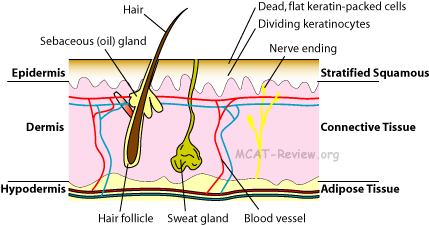
      <div class="fiche-section">
        <div class="fiche-section-title">Fonctions dans la thermorégulation</div>
<ul class="fiche-list">- hairs help insulate the body by trapping air in them.
- Normally hair lies at an angle to the skin, with erectile muscle attaching to it.
- When it's cold, erectile muscles contract, and the hair stands up. This erect position helps hair to trap more air, providing better isolation.
- fat layer for isolation: fat in hypodermis act as isolation.
- sweat glands, location in dermis: produce sweat, cools the body by evaporative cooling.
- vasoconstriction and vasodilatation in surface capillaires
- When it's cold: vasoconstriction of artérioles reduce blood supply to skin capillaires. Leads to less heat loss at skin surface.
- When it's hot: vasodilatation of artérioles increase blood supply to the skin capillaires. Leads to more heat loss at skin surface.
      </div>

      <div class="fiche-section">
        <div class="fiche-section-title">Protection physique</div>
<ul class="fiche-list">- nail = hard keratin = tougher than the soft keratin on skin.
- calluses = extra thick layer of dead keratin-packed cellules on the surface of skin.
- hair = hard keratin.
- protection against abrasion, disease organisms
- Keratin protect skin against abrasion.
- The tight seal made from keratin-packed cellules and glycolipids form a barrier against agents pathogènes.
- Chemical protection: Sweat is acidic, contains anticorps, and antimicrobial agents. Sebum (skin oil) kills bacteria.
- Natural flora: good bacteria on the surface of skin don't cause harm to you, and they fight off bad bacteria that can harm you.
      </div>

      <div class="fiche-section">
        <div class="fiche-section-title">Structure</div>
<ul class="fiche-list">- Epidermis = stratified squamous tissu épithélial = protection
- Keratinocytes = cellules that produce keratin = dominates the epidermis.
- Keratinocytes start off like normal cellules at the bottom of the epidermis, but gets flatter as you go up, and becomes dead, keratin plates at the surface of the skin.
- Melanocytes = cellules that make melanin, the skin pigment.
- Dendritic cellules (Langerhans cellules) = phagocytes that eat agent pathogène and present foreign antigènes to activate réponse immunitaire.
- Dermis = tissu conjonctif = blood, nerf supply
- Fibroblasts = make fiber and ground substance (glue) for the matrice extracellulaire that makes up tissu conjonctif.
- Hair follicles, sweat glands, and oil (sebum) glands.
- Blood vessels and nerfs.
- Hypodermis = tissu adipeux = absorbs shock and provides isolation.
- Review tissu types here
- relative impermeability to water: due to layer of dead, keratin-packed cellules sealed with glycolipids.
- Keratin is water insoluble, and layers of dead, keratin-packed cellules reside on the skin surface.
- Glycolipids seal the space between the dead keratin-packed cellules.
- Sebum (skin oil) contribute some. But oil glands are not present everywhere (absent in palms and soles).
      </div>

      <div class="source-note">Contenu adapté de <a href="https://mcat-review.org/skin-system.php" target="_blank" rel="noopener">mcat-review.org</a> — Traduction et mise en forme par Logopoïos</div>
    </div>
  </div>

  <div class="chapter" id="ch-18">
    <div class="chapter-header" onclick="toggleChapter(this)">
      <div>
        <div class="chapter-num">Chapitre 18</div>
        <div class="chapter-title">Cellules eucaryotes spécialisées et tissus</div>
      </div>
      <div class="chapter-toggle">▼</div>
    </div>
    <div class="chapter-body">

      <div class="fiche-section">
        <div class="fiche-section-title">Cellule nerveuse/Neurone</div>
<ul class="fiche-list">- Contains noyau and organites just like any other cellule.
- Has well-developed RER and golgi (makes a lot of protéines).
- Axone (structure, function)
- Axone = Conducting region of the nerf.
- Axone terminals = secretory regions of nerf.
- Other names for terminaison axonale = bouton synaptique = bouton.
- Dendrites (structure, function)
- Receptive region of the nerf = gets input.
- The branching helps to increase the surface for reception.
- Myelin sheath, cellules de Schwann, oligodendrocytes, isolation of axone
- Myelin sheath = Covers the axone intermittently, with gaps called nœuds de Ranvier.
- The purpose of gaine de myéline is to speed up conduction by insulating the nerf in intervals. This intermittent isolation causes potentiel d'action to jump from one nœud de Ranvier to the next.
- cellules de Schwann = makes gaine de myéline in the système nerveux périphérique by wrapping around the axone.
- Oligodendrocytes = the système nerveux central analogue of cellules de Schwann, makes gaine de myéline around CNS axons.
- Isolation of axone = achieved by the gaine de myéline. Isolation occurs in intervals, which causes potentiel d'action to jump from one nœud de Ranvier to the next.
- Myelin sheath is a good insulator because it is fatty and does not contain any channels.
- Nodes of Ranvier (role in propagation of influx nerveux along axone)
- Action potential jumps from one nœud de Ranvier to the next.
- This jumping of potentiel d'action speeds up conduction in the axone.
- Synapse (site of impulse propagation between cellules)
- Synapse = conduction from one cellule to another.
- Axodendritic synapse = terminaison axonale of one neurone (presynaptic) → dendrite of another neurone (postsynaptic).
- Axosomatic synpase = terminaison axonale of one neurone (presynaptic) → corps cellulaire of another neurone (postsynaptic).
- Axoaxonic synapse (rare) = terminaison axonale of one neurone (presynaptic) → axone hillock of another (postsynaptic).
- Synaptic activity
- transmitter molecules
- Transmitter molecules = neurotransmetteurs
- Action potential → release of neurotransmetteurs by presynaptic terminaison axonale → picked up by récepteur of postsynaptic neurone.
- Release of neurotransmetteur = exocytose of vesicles containing neurotransmetteurs. Triggered by calcium influx when potentiel d'action reaches terminaison axonale.
- Neurotransmetteur reception = diffusion of neurotransmetteur across the fente synaptique, binds to récepteur, opens up ion channels that causes a change in potentiel de membrane of the postsynaptic neurone (potentiel gradué). If this potentiel gradué is large enough, it will trigger a full-fledged, all-or-nothing potentiel d'action in the postsynaptic neurone.
- Neurotransmetteurs are quickly eliminated (destroyed by enzymes, reuptake by presynaptic terminal, or diffuse away) so that they don't persistently stimulate the postsynaptic neurone.
- Neurotransmetteur molecules:
- Acetylcholine (ACh)
- Norepinephrine (NE)
- Dopamine
- Serotonin
- Histamine
- ATP
- boutons synaptiques
- Synaptic knob is another name for terminaison axonale.
- Contains vesicles of neurotransmetteurs waiting to be exocytosed.
- Action potential reaching the bouton synaptique causes an influx of calcium, which signals the vesicles to fuse with membrane cellulaire (exocytose) to release the neurotransmetteurs into the fente synaptique.
- fatigue
- Continuous synaptic activity → depletion of neurotransmetteurs → fatigue.
- propagation between cellules without resistance loss
- Action potential is all-or-nothing.
- As long as the neurotransmetteurs cause the postsynaptic cellule to reach a certain potentiel seuil, the potentiel d'action induced is just as large as the presynaptic potentiel d'action.
- In summary, propagation between cellules involves no resistance loss because the postsynaptic potentiel d'action is just as large as the presynaptic potential - all potentiel d'actions are all-or-nothing.
- Resting potential (gradient électrochimique)
- Na+-K+ pump = 3 Na+ out, 2 K+ in = net negative to the inside, net positive to the outside.
- K+ leakage = the resting membrane cellulaire has channels that allow K+ to leak out, but don't allow Na+ to leak in = net negative to the inside, net positive to the outside.
- Resting potential is -70 mV because the cellule is more negative on the inside, and more positive on the outside.
- Electrochemical gradient = combination of electrical and chemical gradient = both electrical potential and ion gradient de concentration across membrane.
- Action potential
- Stages of an potentiel d'action:
- Resting: cellule at rest, sodium-potassium pump maintaining potentiel de repos (-70 mV). Lots of sodium outside, lots of potassium inside. Ion channels closed so the established ion gradient won't leak.
- Dépolarisation: sodium channels open, positive sodium rushes inside, potentiel de membrane shoots up to +30 mV. Lots of sodium inside, lots of potassium inside.
- Repolarisation: potassium channels open, sodium channels close, positive potassium rushes outside, potentiel de membrane drops back down. Lots of sodium inside, lots of potassium outside (opposite of the resting state).
- Hyperpolarisation: potassium channels doesn't close fast enough, so the potentiel de membrane actually drops below the potentiel de repos for a bit.
- Refractory period: the sodium-potassium pump works to re-establish the original resting state (more potassium inside, sodium outside). Until this is done, the neurone can't generate another potentiel d'action. Absolute refractory period = from dépolarisation to the cellule having re-established the original resting state. Relative refractory period = After hyperpolarisation till resting state re-established.
- threshold, all-or-none
- When a stimulus (potentiel gradué) depolarizes above a threshold value, an potentiel d'action will occur.
- Action potentials are all-or-none, meaning that if it occurs, all potentiel d'action have the same magnitude.
- One potentiel gradué just barely makes the threshold value, another overshoots it a lot, but both will cause the same potentiel d'action.
- sodium-potassium pump
- 3 sodium out.
- 2 potassium in.
- net positive out.
- causes membrane to be more negative on the inside, hence negative potentiel de membrane.
- Excitatory and inhibitory nerf fibers (sommation, frequency of firing)
- Excitatory = stimulates an potentiel d'action to occur
- Excitatory synapse = récepteur binding causes postsynaptic potential to be more positive (dépolarisation) = if it gets above threshold, potentiel d'action results.
- Inhibitory = inhibits an potentiel d'action from occuring.
- Inhibitory synapse = récepteur binding causes postsynaptic potential to be more negative (hyperpolarisation) = makes it more difficult to reach threshold.
- Sommation = two or more nerfs firing at the same time.
- Two subthreshold excitatory nerfs firing at the same time can sum to reach the threshold.
- A threshold excitatory nerf and an inhibitory nerf firing at the same time, and the resultant signal won't reach the threshold.
- Frequency = Firing, then quickly firing again.
- If the first fire is subthreshold, fire again before the previous dépolarisation dies, and the new dépolarisation will be even higher than the first time.
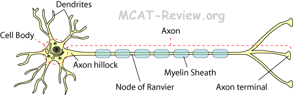
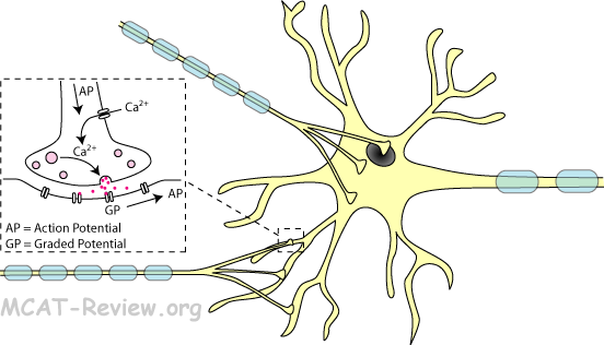
      <div class="fiche-section">
        <div class="fiche-section-title">Cellule musculaire/Contractile</div>
<ul class="fiche-list">- Striated = muscles squelettiques, voluntary, has stripes, multiple nuclei shared within the same muscle fiber. Strong, but tire easily = shaped like long fibers.
- Smooth = visceral, involuntary muscles, no stripes, single noyau per cellule. Weak, but doesn't tire easily = shaped like almonds, tapered on both ends.
- Cardiac = muscle cardiaques, involuntary, has stripes, single noyau per cellule, strong and doesn't tire easily = highly branched, shaped like fibers cross-linked to one another.
- Abundant mitochondries in red muscle cellules (ATP source)
- Red muscle = high endurance, but slow.
- Aerobic respiration predominant.
- Many mitochondries because red muscles undergo respiration aérobie.
- Equipped to receive abundant apport en oxygène: many capillaires, many myoglobin.
- High endurace, doesn't tire easily.
- White muscle = fast, but fatigue easily.
- Anrespiration aérobie (glycolyse) predominant.
- Few mitochondries because white muscles undergo mainly glycolyse.
- Equipped for short bursts of glycolyse: stores high amounts of glycogen.
- Pink muscle = intermediate between red and white muscle.
- Organization of contractile elements (actin and myosin filaments, ponts transversaux, modèle du filament glissant)
- Actin filament = filament fin = has troponin and tropomyosin on it.
- Myosin filament = filament épais = has myosin heads on it.
- Cross bridge = myosin head binds to actin.
- Sliding filament model = Cross bridge forms, myosin head bends (coup de force), causes actin to move (slide) in the direction of the coup de force (toward the M line). When all the actin slide toward the M line like this, the muscle fiber contracts.
- Something counter-intuitive about the modèle du filament glissant: ATP is not directly needed for the powerstroke. ATP binding is needed for detachment of myosin head to actin. ATP hydrolysis is needed for de-powerstroke (unbend myosin head).
- Rigor mortis = no ATP after a person dies, myosin heads can't detach after coup de force, muscle remain in contracted position.
- So what is troponin and tropomyosin there for? Ans: tropomyosin on actin blocks the myosin head from forming ponts transversaux. However, troponin moves tropomyosin out of the way at high Ca2+ levels (Ca2+ binds to troponin, and troponin moves tropomyosin).
- Calcium régulation of contraction, réticulum sarcoplasmique
- Sarcoplasmic reticulum (SR) = RE lisse in muscle = stores calcium, releases them in response to AP.
- The SR is also called terminal cisternae where it meets T-tubules at the edges of the sarcomere.
- T-tubule = extension of the muscle membrane cellulaire that runs deep into the cellule, so that potentiel d'action can reach there.
- Muscle contraction:
- Nerf stimulates muscle.
- Action potential runs along muscle membrane cellulaire.
- Goes deep into the muscle cellule via T-tubules.
- Stimulates the SR (terminal cisternae) to release calcium.
- Calcium causes muscle to contract via the mécanisme du filament glissant.
- Sarcomeres ("I" and "A" bands, "M" and "Z" lines, "H" zone - General structure only)
- I band = thinnest = filaments fins only = sides of the sarcomere.
- H zone = fattest = filaments épais only = center of the sarcomere, spans the M line.
- A band = contains both thick and filaments fins, center of the sarcomere spans the H zone.
- M line = line of myosin in the middle of the sarcomere, linked by accessory protéines.
- Z line = zigzag line on the sides of the sarcomere, connects the filaments of adjacent sarcomeres.
- mnemonics
- I = thin like the letter I.
- H = fat like the letter H.
- A = letter width in between I and H, so a mixture of thick and filaments fins.
- M = middle line = myosin (linked by accessory protéines).
- Z = zigzag line
- Moving from middle to the side of sarcomere = M HAIZ, the Muscle says HAIZ.
- Presence of troponin and tropomyosin
- Tropomyosin = long protéine that spirals along actin, blocks myosin head from cross-linking.
- Troponin = binds tropomyosin, moves it out of the way when calcium is around.
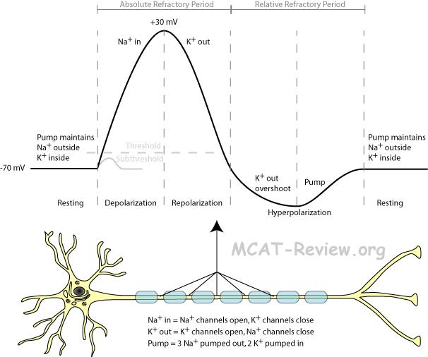
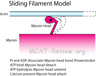
      <div class="fiche-section">
        <div class="fiche-section-title">Autres types de cellules spécialisées</div>
<ul class="fiche-list">- Squamous = flat.
- Cuboidal = cube.
- Columnar = column shaped.
- Simple epithelium = single cellule layer = good for absorption, sécrétion, filtration, diffusion.
- Simple squamous: endothelium, capillaire wall, alvéolaire wall.
- Simple cuboidal: gland ducts, rein tubules.
- Simple columnar: estomac and gut.
- Stratified epithelium = two or more cellule layers = good for protection against abrasion.
- Stratified squamous: skin.
- Stratified cuboidal/columnar: not common.
- Endothelial cellules
- Endothelial cellules = lines the inside of organes and vaisseaux sanguins = simple squamous epithelium.
- Thin, single layer cellules facilitate diffusion.
- Connective tissu cellules (major types cellulaires, fiber types, loose vs. dense, cartilage, matrice extracellulaire)
- Connective tissu structure = Cellules + matrice extracellulaire.
- Cellules: secrete the matrice extracellulaire (ground substance and fibers).
- Ground substance: glue that holds the matrix together.
- Fibers: mostly collagen, gives the matrix strength.
- Connective tissu cellules and tissu types: bone, fat, tendons, ligaments, cartilage, blood.
- Osteoblasts make bone.
- Fibroblasts make tissu conjonctif proper (fats, tendons, ligaments, beneath epithelia).
- Chondroblasts make cartilage.
- Hematopoietic cellules souches make blood.
- Nomenclature:
- -blast = cellule souche actively producing matrix.
- -cyte = mature cellule, doing housekeeping.
- Fiber types:
- Collagen = the most common fiber type. Very strong. Present in large amounts in dense tissu conjonctif.
- Elastic fibers = can stretch.
- Reticular fibers = can branch and form nets. Found in loose tissu conjonctif.
- loose vs. dense
- Loose = loose fibers, lots of fluff (ground substance, cellules) = anything that you don't associate with being fibrous = fat, paddings around organes.
- Dense = dense fibers predominantly collagen = genuinely fibrous, little fluff (ground substance, cellules) = tendon, ligament.
- Cartilage = chondrocytes + matrix = elastic, flexible, used as padding in spinal discs, ends of bones, ear.
- Extracellular matrix = secreted by cellules = ground substance (glue) and fibers.
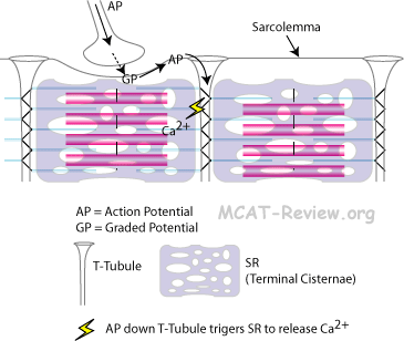
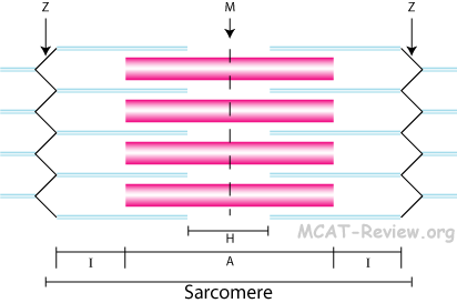
      <div class="source-note">Contenu adapté de <a href="https://mcat-review.org/specialized-eukaryotic-cells-tissues.php" target="_blank" rel="noopener">mcat-review.org</a> — Traduction et mise en forme par Logopoïos</div>
    </div>
  </div>

</div>